Fisioterapia e Riabilitazione del Ginocchio a Roma | Fisio Point
Fisio Point Centri di fisioterapia, poliambulatorio, diagnostica, risonanza magnetica a Roma, Monteverde, Eur Torrino, Trionfale e Grottaferrata_fisioterapista

Knee Physiotherapy and Rehabilitation in Rome

If you are looking for a Physiotherapy and Knee Rehabilitation Centre in Rome, rely on Fisio Point.

The knee is one of the joints most susceptible to direct and indirect trauma, and is greatly affected by postural and walking changes that inevitably lead to premature wear and tear of the knee structures. The knee is an extremely mobile and complex joint, which connects three bones: the femur, the tibia and the patella via the patellofemoral joint and the patellofemoral joint.

La patella is a flat bone contained in the tendon of the quadriceps muscle, which plays a fundamental role in the knee, both because it is the fulcrum in the transmission of force during extension of the leg, and because it provides protection for the internal structures especially when the knee is bent.

Within the knee joint we have two cartilage structures of fundamental importance: the medial meniscus and the meniscus side. The menisci are connected by the intermeniscal ligamentand attached to the tibia via the anterior meniscal ligaments e rear. The main role of the menisci is to ensure congruence between the articular surfaces and mechanical protection from the stress to which the knee is subjected.


Book a Free Visit

Do you want to understand the origin of your problem and structure the best physiotherapy path to solve it definitively?

If you have them, bring us medical prescriptions, MRIs and X-rays. This way we can assess the situation together and determine the best course of action to deal with your problem and resolve it quickly. If you do not have them and they are necessary, we can do them quickly ourselves.

General (#17)

Fill in the form,


You will be contacted as soon as possible


General (#17)

Fill in the form,


You will be contacted as soon as possible


The articular surfaces of the knee are covered by cartilage, a special fabric that protects the bones by reducing internal friction during movement. 

As with any articulation of this type, there is a capsule articulate which surrounds the entire joint and secretes a lubricating liquid called liquid synovial which promotes joint mobility by preventing friction and nourishing the area.

Of fundamental importance for both static and dynamic knee stability are the ligaments. The main ligaments of the knee are: the anterior cruciate ligament (lca) and the ligament crusader rear (lcp) which originate from the base of the femur and insert on the tibial plateau. Their function is to limit the rotation and antero-posterior movement of the tibia in relation to the femur. Supporting the lateral parts of the joint are the ligament collateral medial (lcm) or internal collateral and the external collateral ligament or collateral side (lcl), which join the femur with the tibia and the femur with the fibula head respectively.

In addition to these, which are the main ligaments, there are others such as

  • the patellar ligament, linking the patella (kneecap) with the tibia
  • i oblique and arcuate popliteal ligaments, that provide stability at the back of the knee.

Knee stability is also guaranteed by other structures that are the tendons and the muscles. Very important is the role of the quadriceps, the largest muscle of the thigh, which allows all extension movements and inserts on the tibia forming the patellar tendon. As for the dynamic stabilisers, medially we have the tendon of the semimembranosus muscle, and laterally the biceps femoris and popliteus muscles.

We often hear about 'goose foot' tendons, which is the structure formed by the tendons of three muscles: frail, sartorius e semitendinosus. They are located on the inner surface of the tibia, separated from the medial collateral ligament by a serous bursa. Inside the knee there are also small fluid-filled sacs called 'bursae', such as the bursa prerotulae and the infraotulae, which are located between two tendons or between a bone and a tendon and whose function is to protect the structures from excessive stress and friction.

Fisio Point, fisioterapia roma, poliambulatorio, idrokinesi, risonanza magnetica Roma, Monteverde, Eur, Grottaferrata. Tecar terapia

Knee Diseases and Treatments

Meniscus Injury

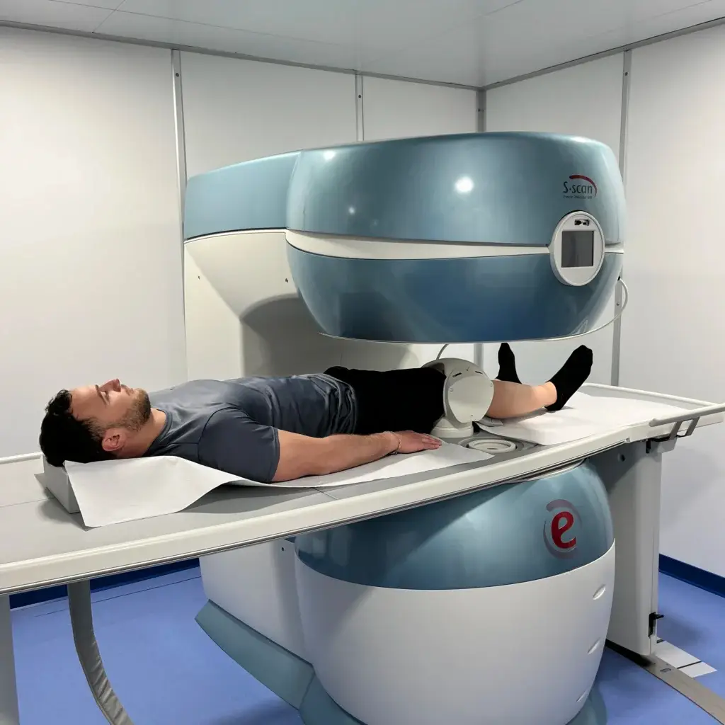
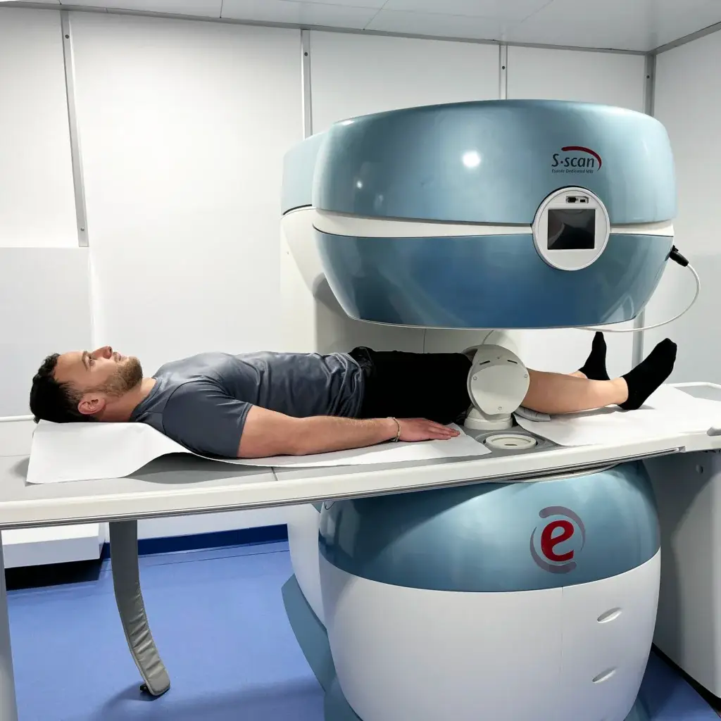
The meniscusespecially because of its role within the knee and its location, is often prone to injury. Posture and load distribution play a key role. Remember that on menisciwe always load the entire weight of our body up to the kneeare therefore particularly high-risk facilities. Statistically, most injuries of a traumatic nature occur when the knee is in torsion. A meniscus broken causes localised pain, often the knee swells and in some cases, if fragments of meniscus move within the joint, it can even be blocked. In case of trauma or suspected breaking meniscal it is a good idea to have an MRI scan to see precisely the extent of the damage.

Meniscus Injury Treatment

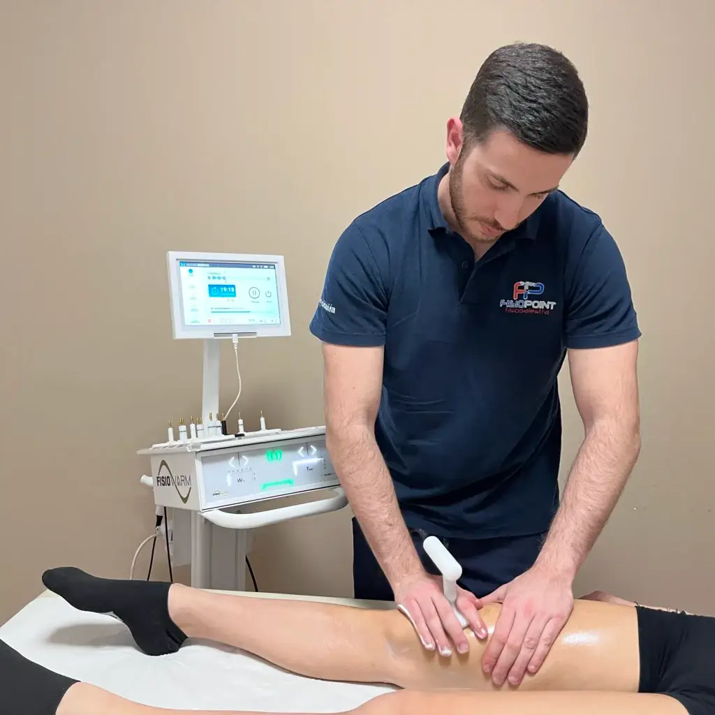
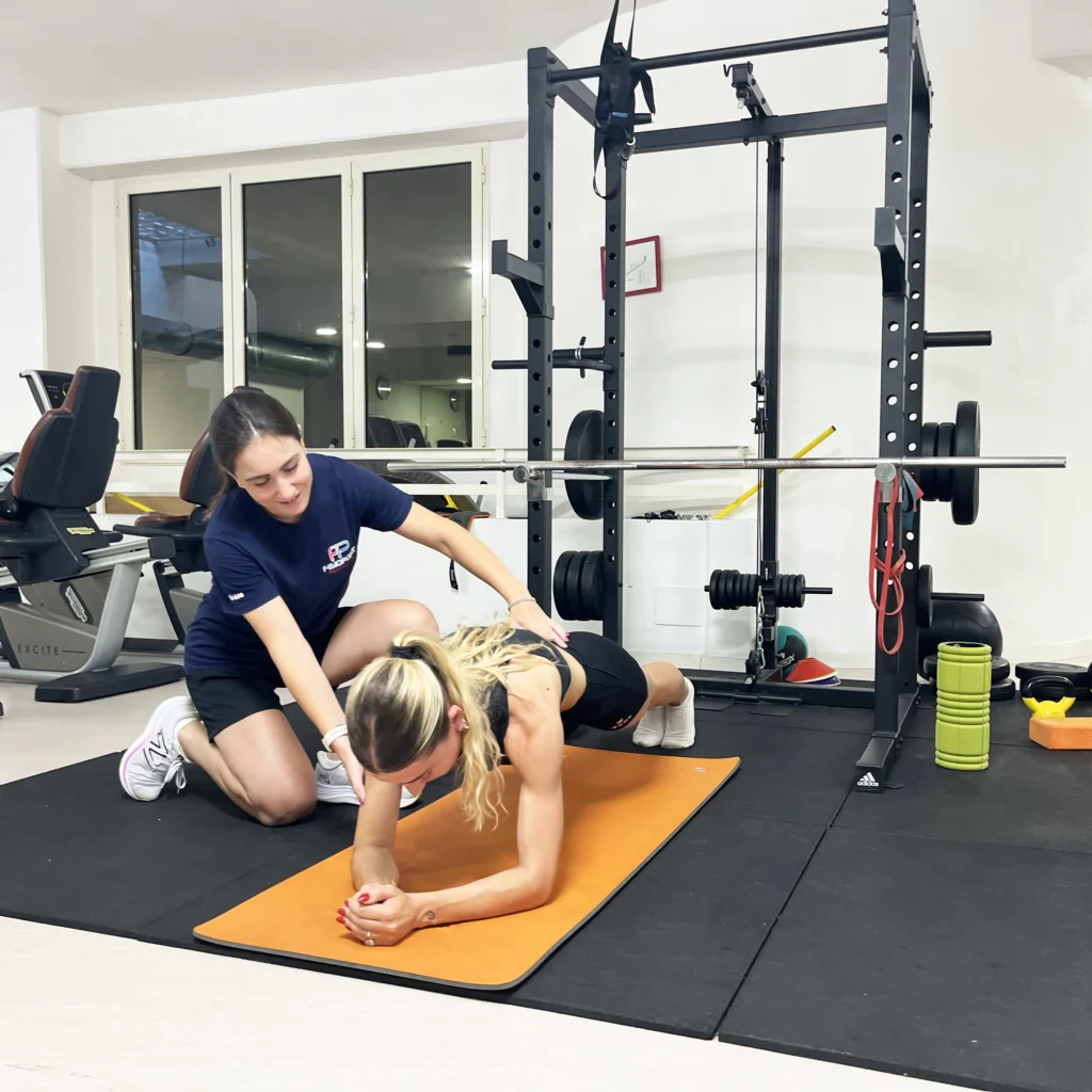
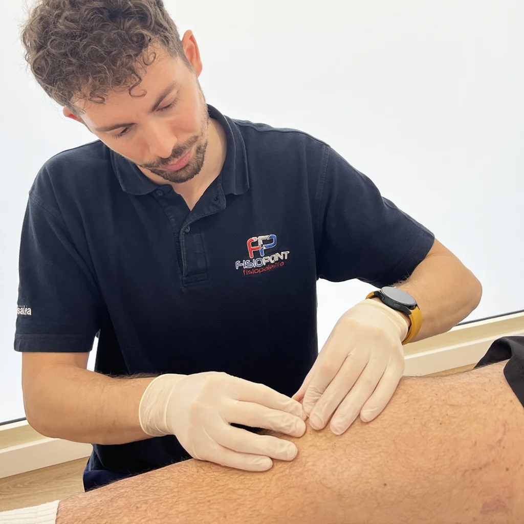
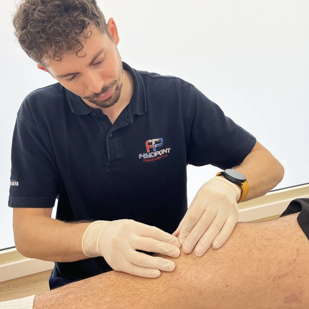
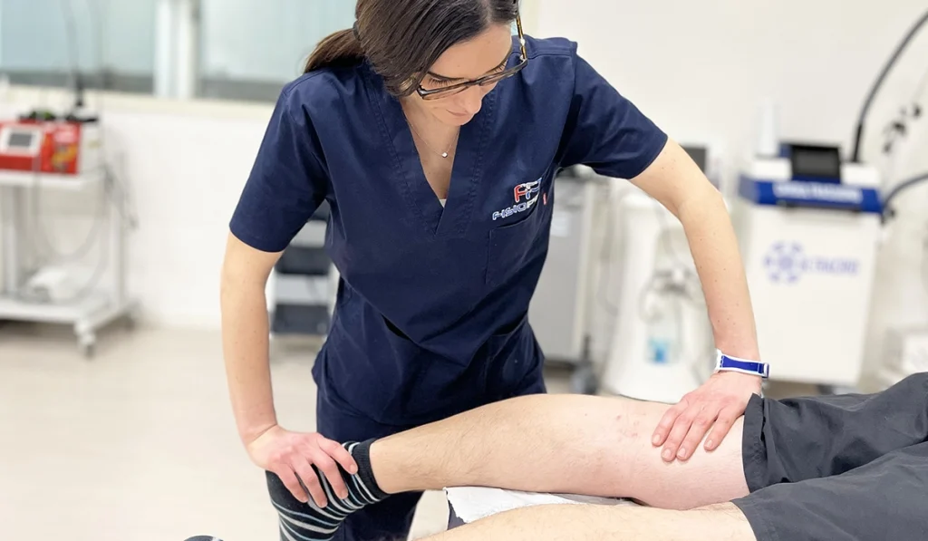
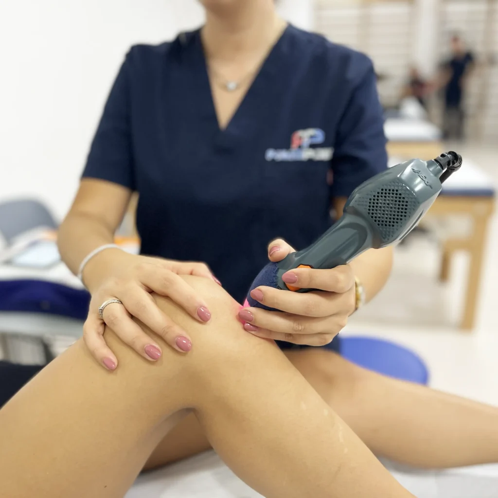
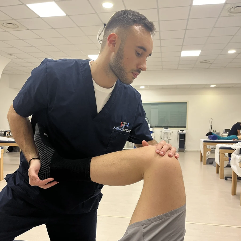
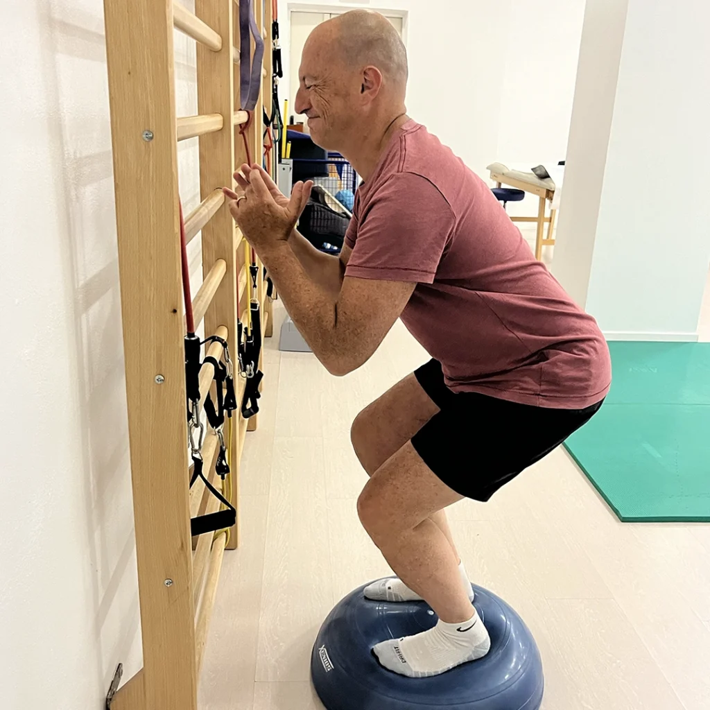
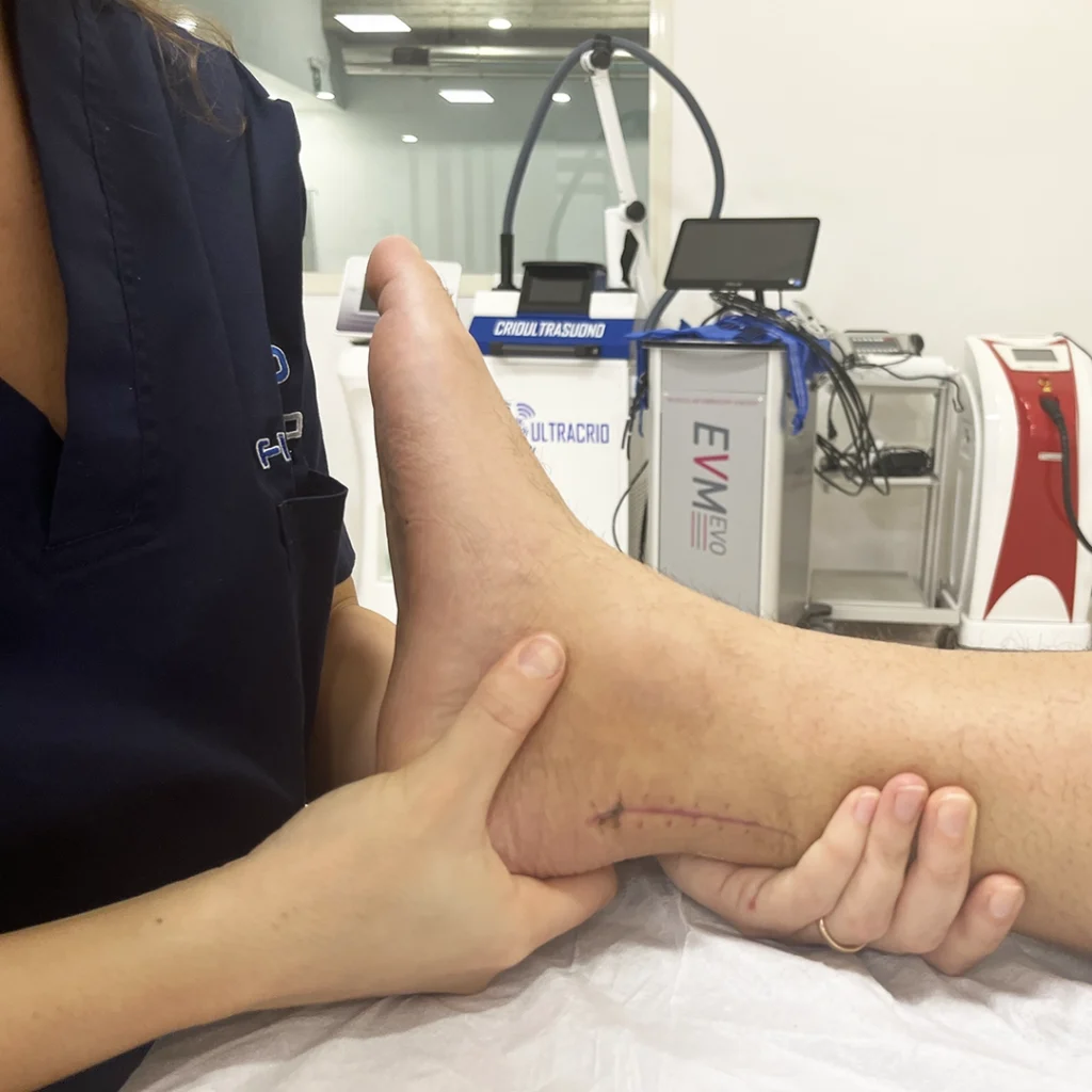
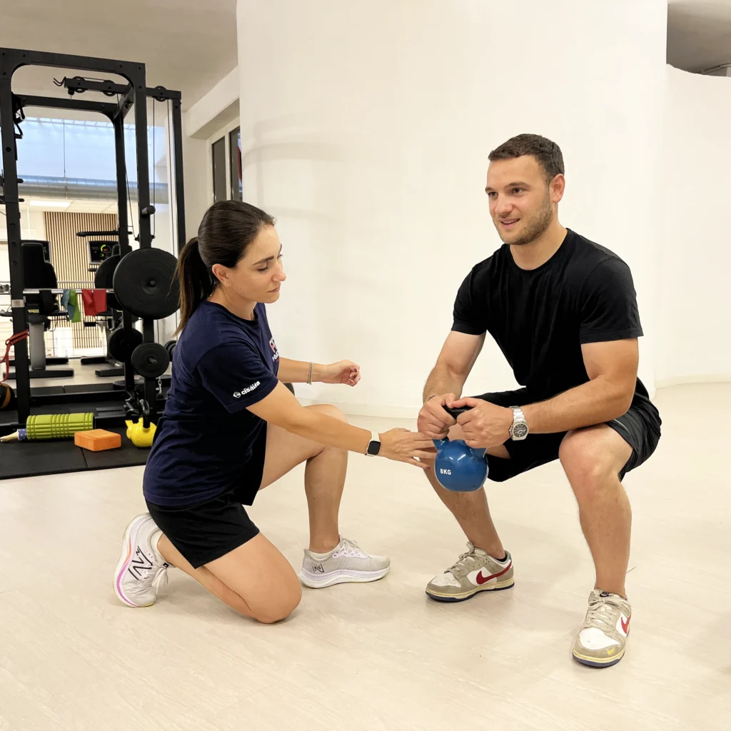
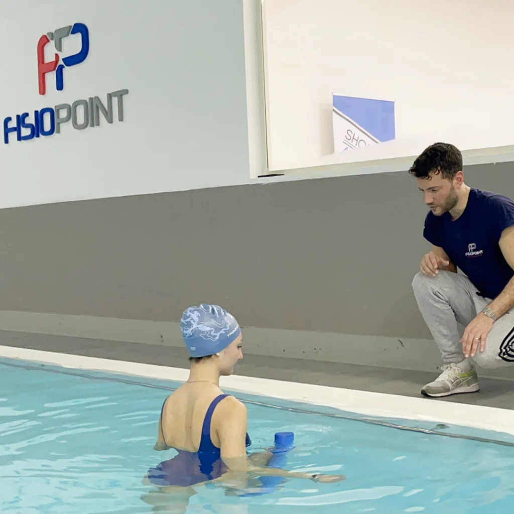
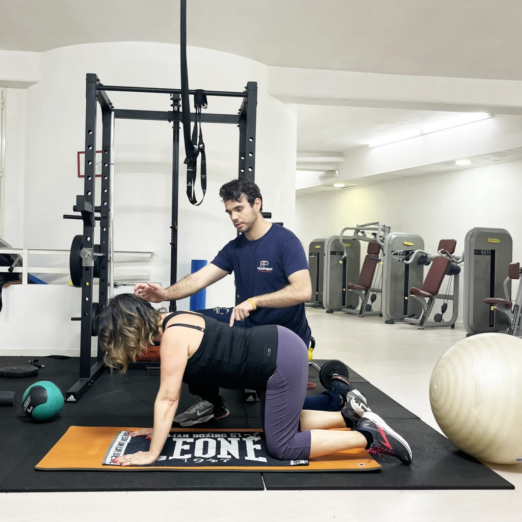
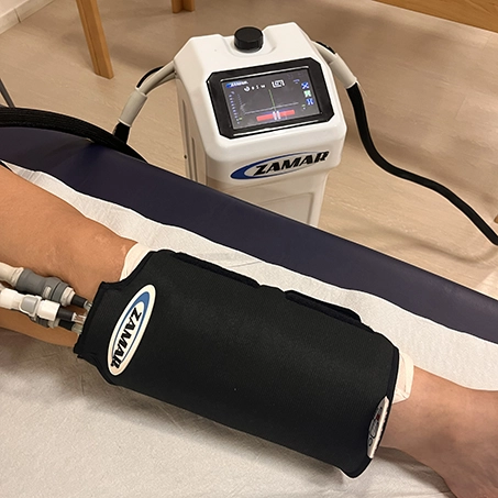
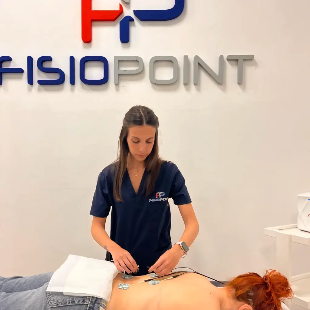
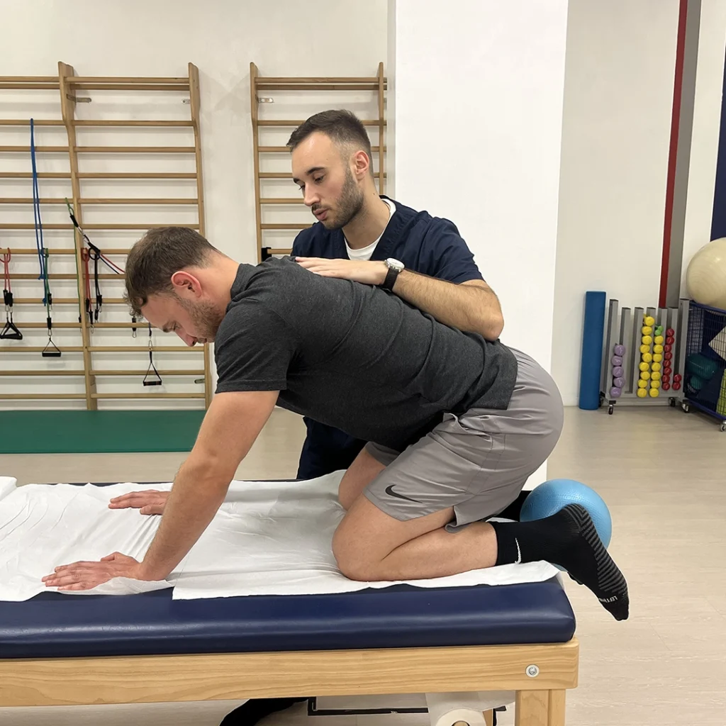
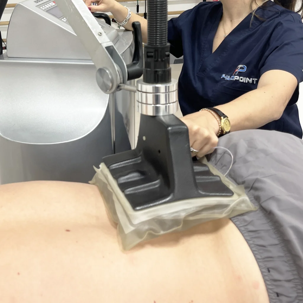
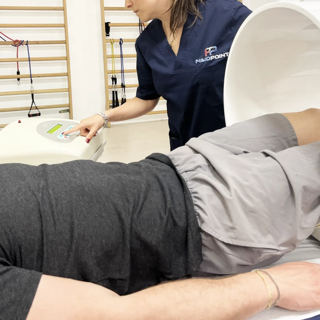
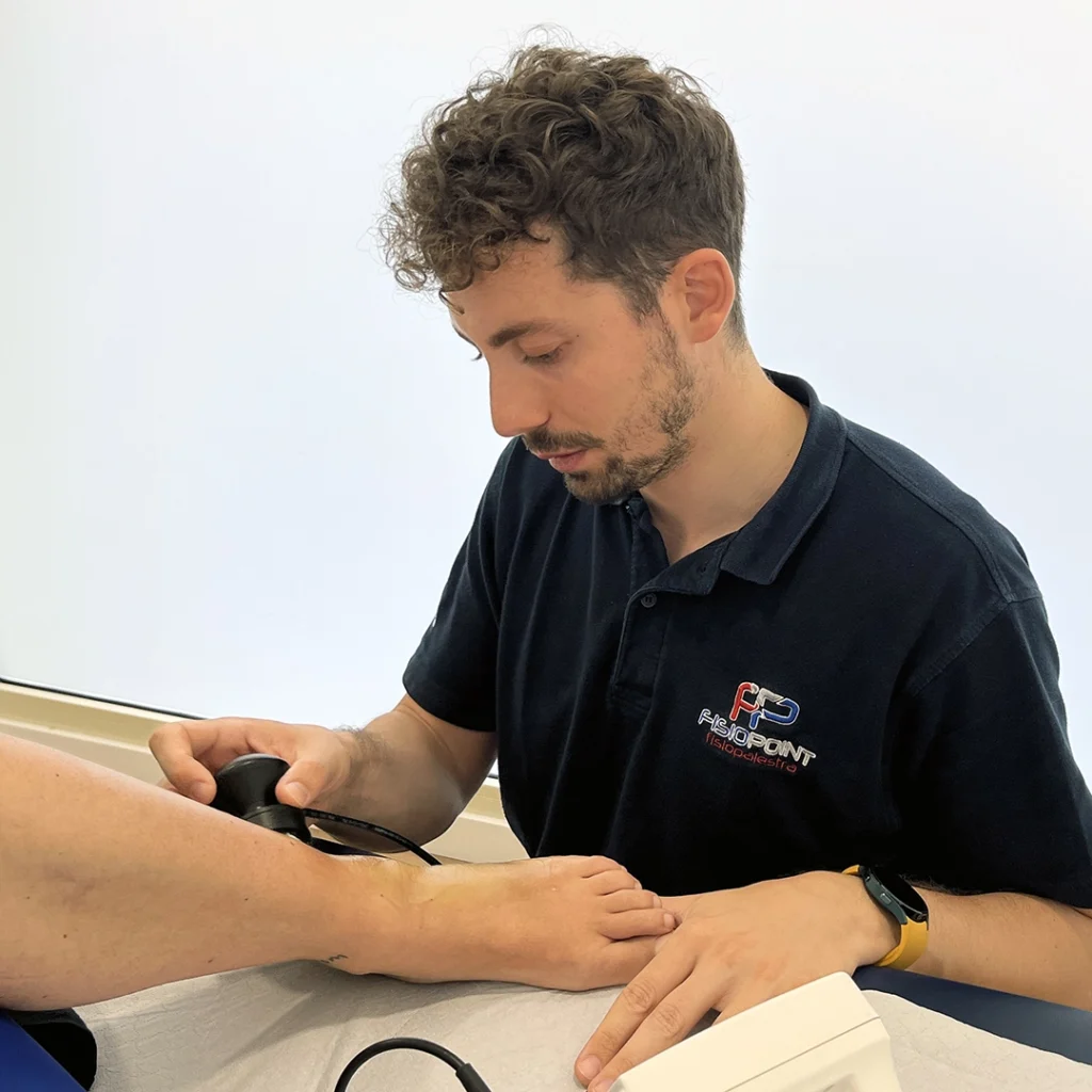
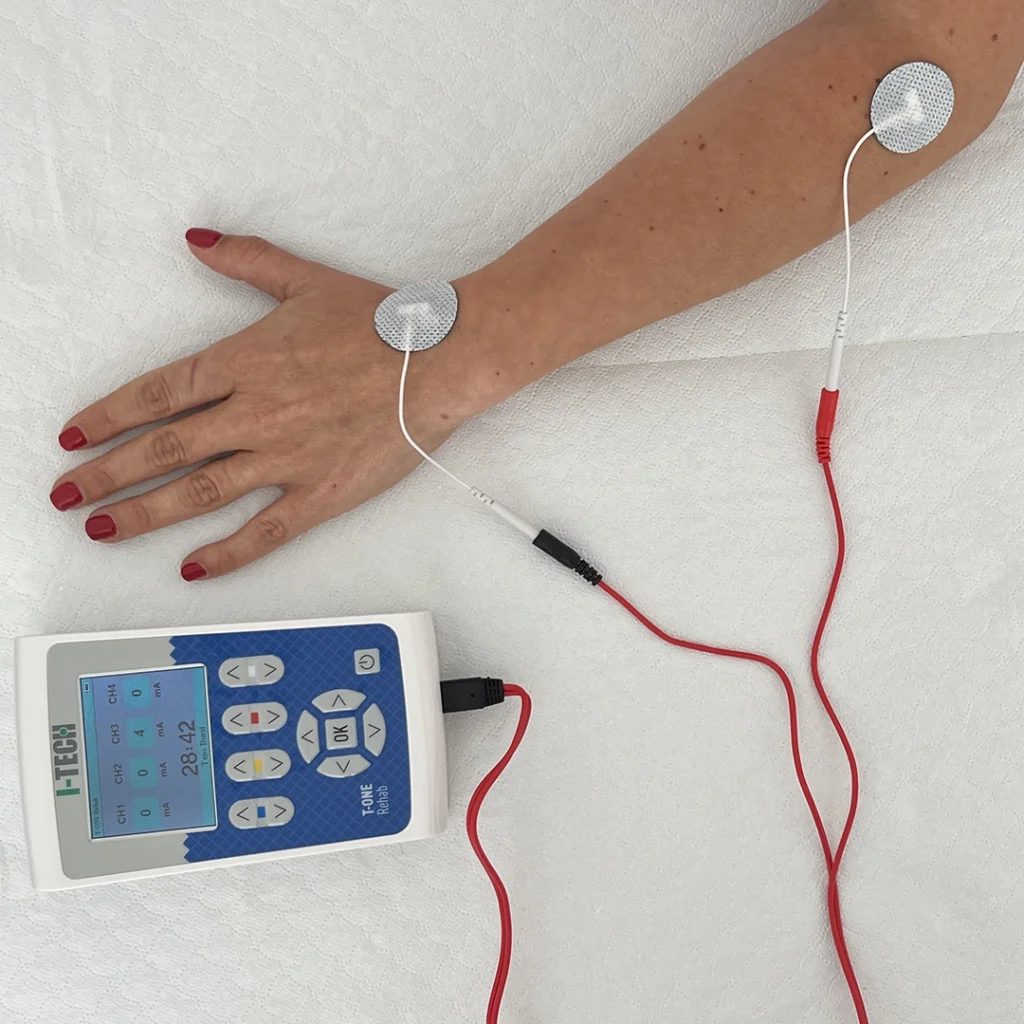
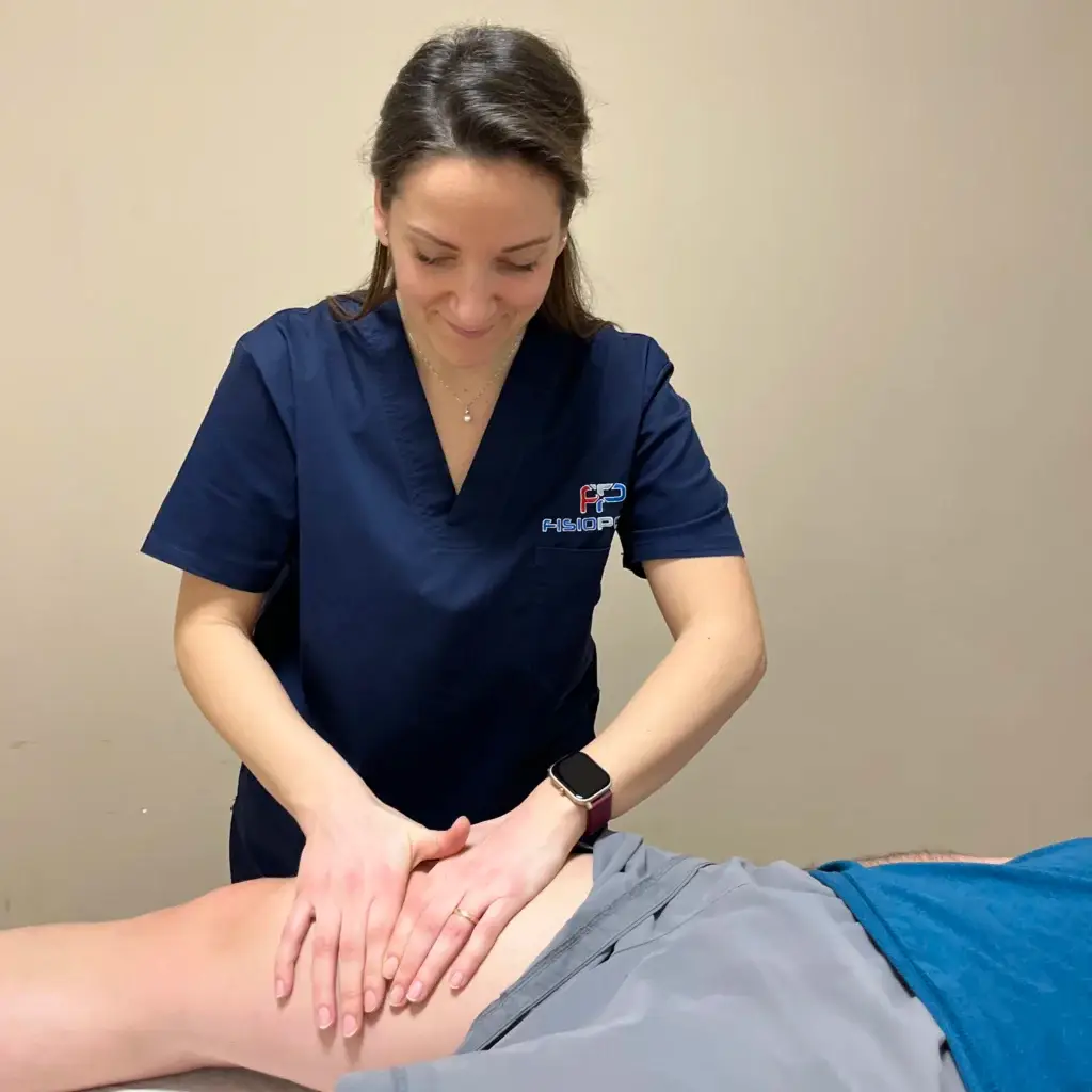
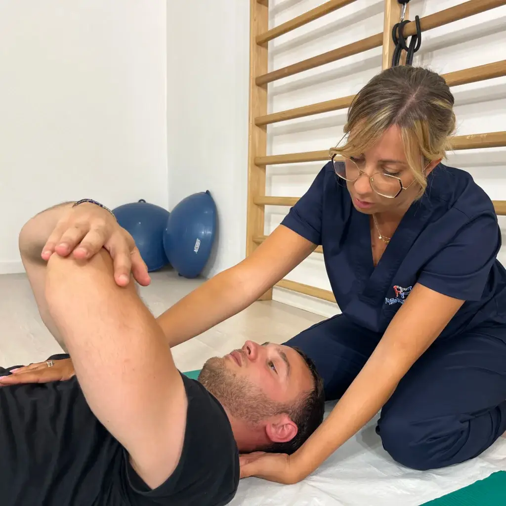
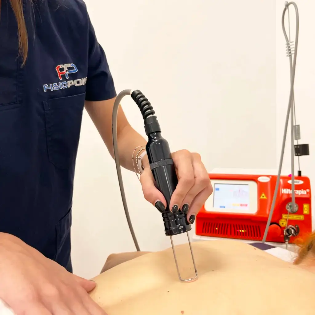
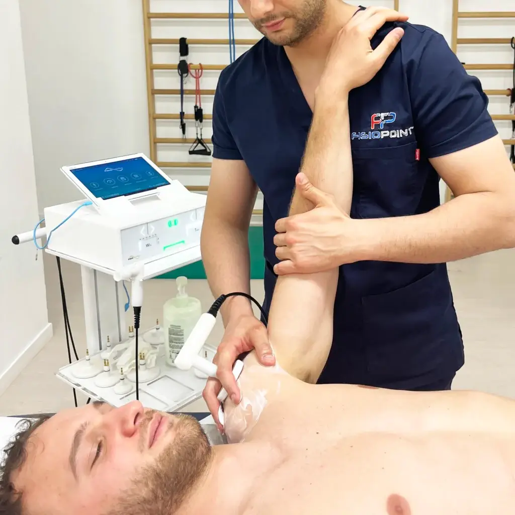
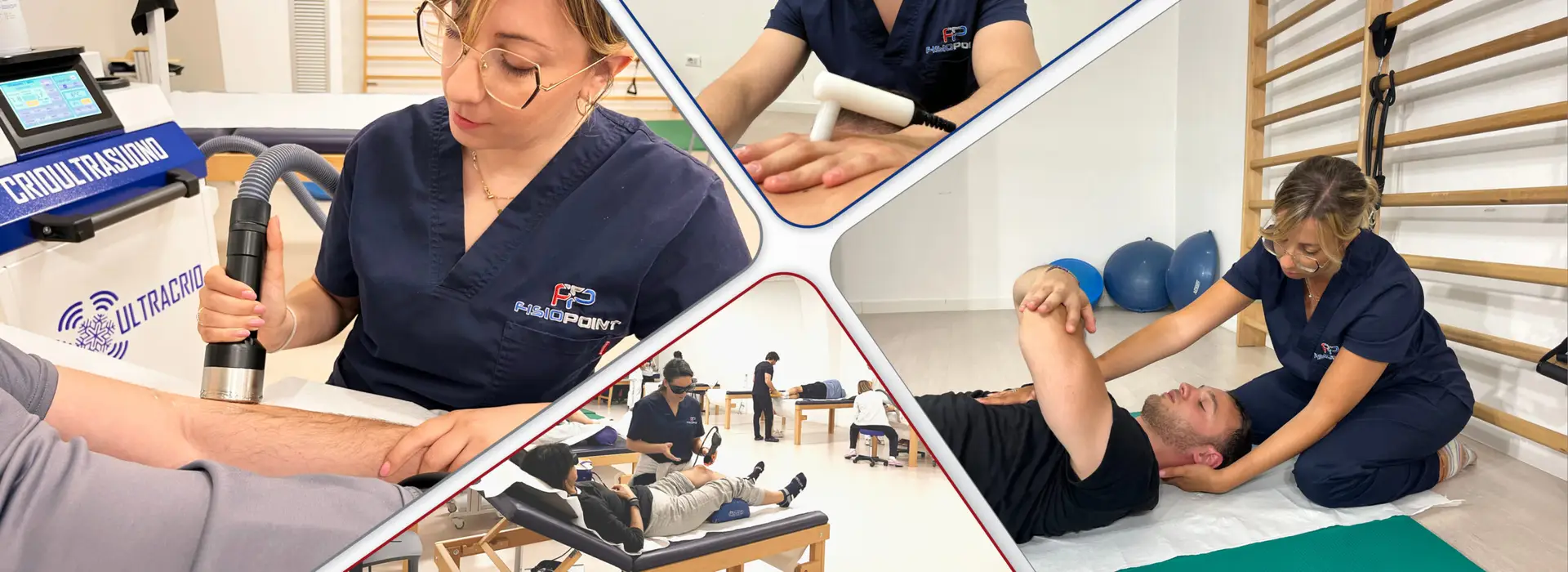
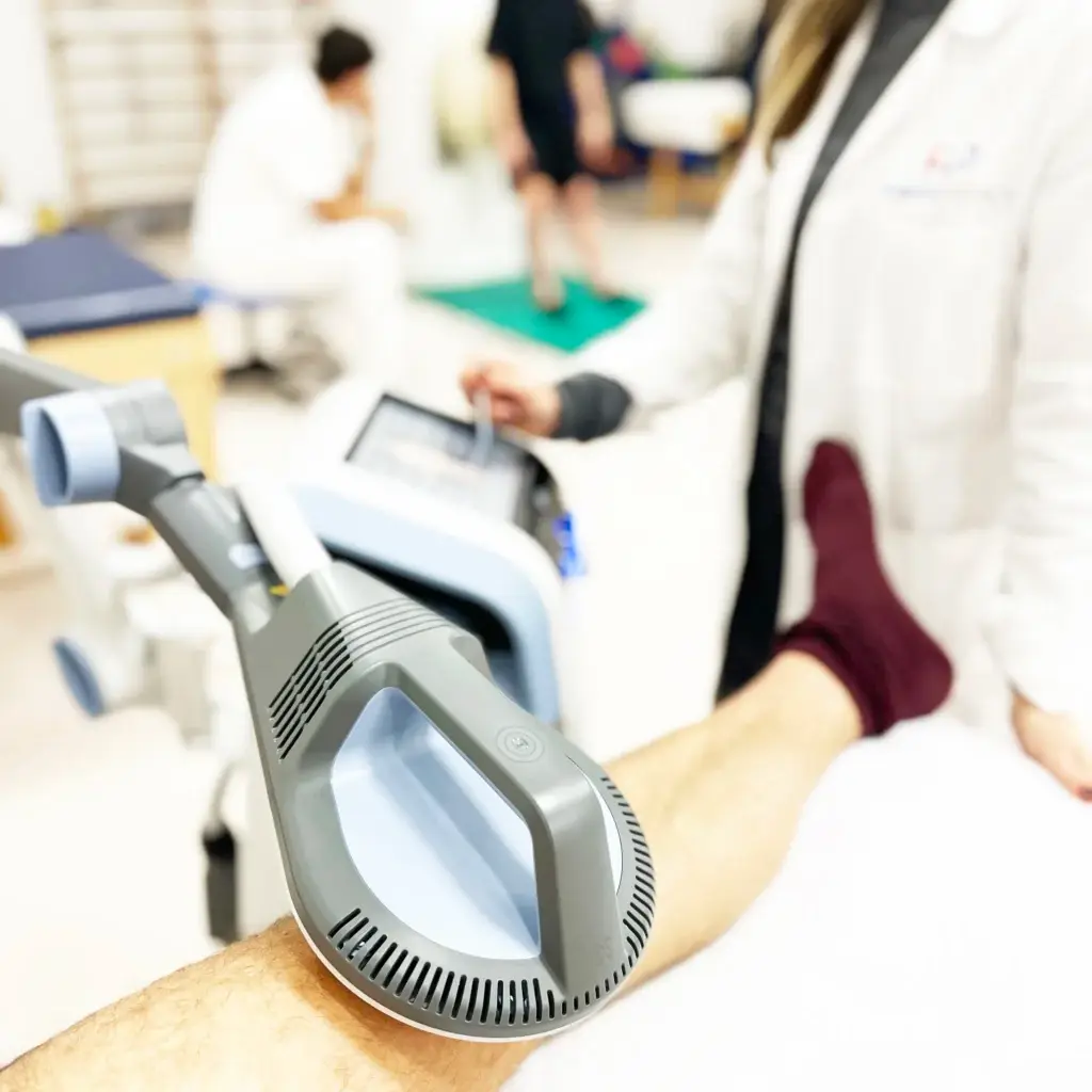
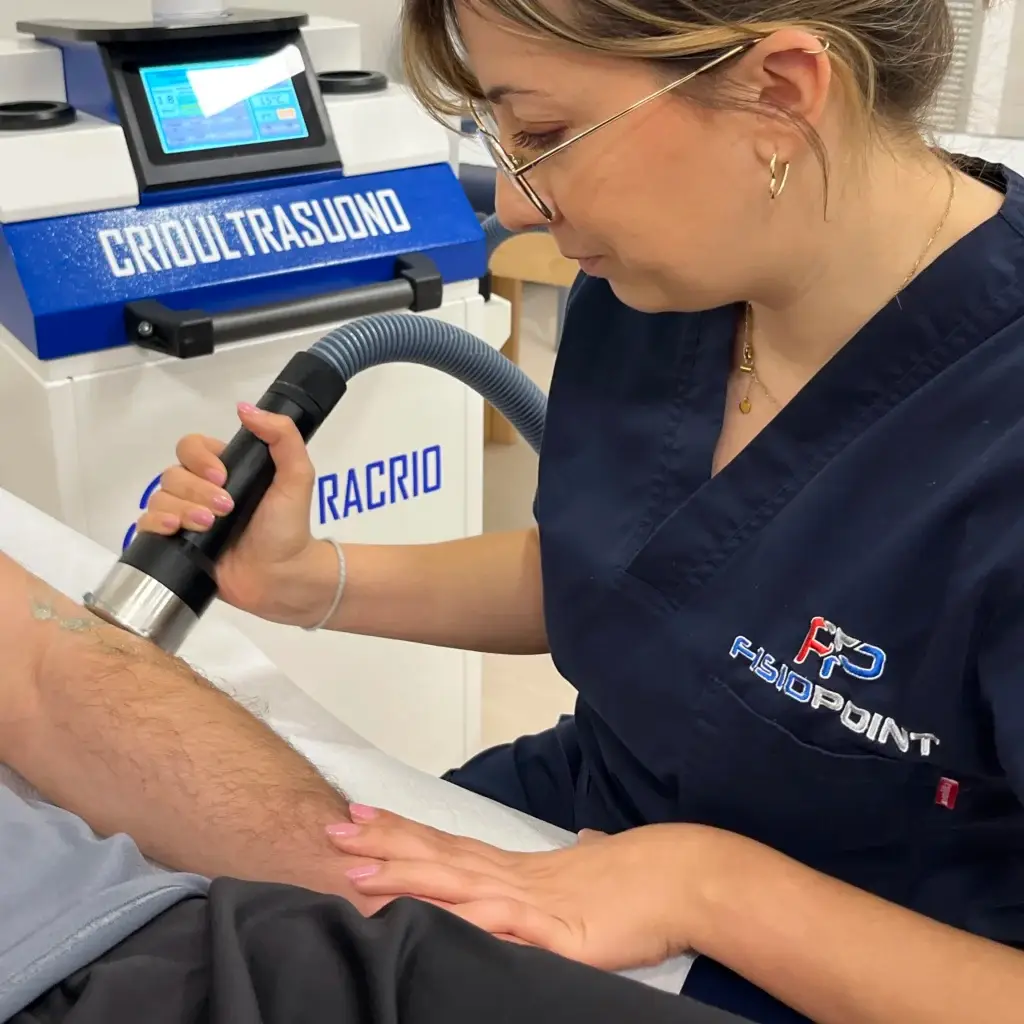
Depending on the extent of the injury of meniscus and the general situation of the knee one can proceed conservatively by doing adequate physiotherapy with the most modern instrumental therapies such as the Tecar therapy, the Yag laser high power, associated with Hydrokinesiotherapymanual treatments, proprioceptive exercises and strengthening exercises. Initially the goal is to de-injure, remove pain and recover articularity, then the work aims at recovering strength and proprioceptive control and stability.

When the meniscus rupture is such that it cannot be resolved conservatively, it is necessary to resort to an operation that is nowadays done arthroscopically to suture or remove the broken fragments. After the operation, however, physiotherapeutic treatment is necessary, which is similar to conservative treatment but is longer in some cases.

Anterior, posterior and collateral cruciate ligament injury

Injuries to the anterior cruciate ligament, which are more frequent, or to the posterior cruciate ligament of the knee almost always occur due to trauma of a distortional nature. Often, rupture of one or both cruciate ligaments is also associated with involvement of the collateral ligaments and menisci.

Symptoms are characterised by

  • localised pain
  • functional impotence
  • feeling of knee failure
  • swelling due to an effusion inside the joint.

With these symptoms, it is likely that there is even a partial rupture of the anterior cruciate ligament. In some cases, the extent of the immediate pain does not always correlate with the severity of the cruciate ligament rupture. Often the patient may tell the doctor phrases such as 'I felt my knee move' or 'I felt a tear or a crack'. In any case, it is necessary to ascertain the damage through MRI and an orthopaedic check-up.

LCA treatment

In the meantime, it is necessary to ascertain the severity (degree) of the injury. If the injury is partial, conservative therapy and a period of physiotherapy may be sufficient to restore knee function. In more severe cases, surgery is necessary to reconstruct the affected ligament. Collateral ligaments have a good recovery capacity, so in many cases surgery is not necessary; good physiotherapy with the best instrumental therapies is sufficient. In anterior cruciate ligament (ACL) or posterior cruciate ligament (PCL) injuries, surgery replaces the ruptured ligament using other structures of the knee itself, such as the patellar tendon or the semitendinosus and gracilis tendons, or in some cases using a donor tendon. Depending on the injury and the type of surgery, a brace will be required in the days following the operation and you will be able to walk with the aid of crutches for 10-20 days. In some cases, a Kinetec is recommended to move the knee passively by gradually increasing degrees of flexion. Right from the first few days, it is necessary to rely on an excellent Knee Physiotherapy and Rehabilitation Centre in Rome for good physiotherapy that provides excellent instrumental therapies such as the Tecar therapyhigh-powered Yag laser... which serve to disinfect the knee, take away the pain and reduce the effusion. At the same time, manual work by an experienced physiotherapist on the muscles and the joint is necessary to recover the movement of the knee and begin strengthening exercises to rebuild the muscles that will serve to give stability to the joint. As soon as the stitches are removed, i.e. about 15-20 days after surgery, one can enter the water to do hydrokinesitherapy, which greatly facilitates and accelerates the recovery of movement and strength. A very important aspect in ACL rehabilitation is also the recovery of proprioception, i.e. the control ability that gives balance and stability to the knee.
Book a Free Visit

Contact

Fill in the form,

You will be contacted as soon as possible


Arthrosis of the Knee

Osteoarthrosis is a degenerative process affecting the cartilages that line the articular surfaces of bones, leading to friction between them. It is a process that advances with age, but can be accelerated in some cases by incorrect posture or certain skeletal pathologies such as

  • the varus or valgus knee
  • fractures
  • ligament ruptures that cause knee instability

The most frequent symptoms are

  • restriction of mobility
  • feeling of joint and muscle stiffness
  • more or less localised pain depending on the degree of arthrosis

Arthrosis is a condition that tends to worsen, especially if not treated in time.

Arthrosis treatment

Prevention from the main risk factors, such as being overweight or muscular and ligamentous imbalances that lead to increased strain on the joints, is certainly very important. From the very first symptoms, it is necessary to rely on a qualified Knee Physiotherapy and Rehabilitation Centre in Rome with the most modern instrumental therapies such as the Tecar therapy or the High-power Yag laserassociated with theHydrokinesiotherapy and manual treatments for the recovery of articularity and stability, are able to counteract and significantly improve pain and joint limitation due to arthrosis. In some cases, infiltrations of hyaluronic acid or growth factors are appropriate to 'lubricate' the joint and facilitate recovery. In the most severe cases, an operation involving the insertion of prostheses of varying size depending on the extent of the damage must be performed. Afterwards, a period of rehabilitation and motor re-education is necessary to restore the joint to an optimal condition.
Varo/Valgo Knee

Knee valgus and varus are two of the main knee paramorphisms. It is the condition in which the femur and tibia are not perfectly aligned and form angles, with either an internal (varus knee) or external (valgus knee) opening. There is often no exact cause for the occurrence of these problems and most often the problem arises at a young age. Often a condition of laxity or retraction of the leg musculature can alter the axis of the leg, causing a varus knee or a valgus knee. Other causes are due to neurological, ligament or skeletal problems.

Varo/Valgo knee treatment

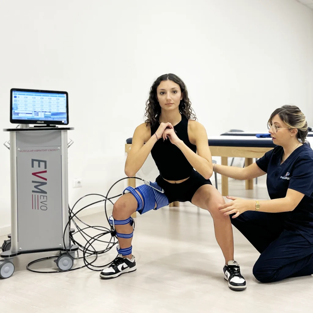
For the correction of valgism and varus of the knee, it is first necessary to make an assessment of the posture, a baropodometry to see the static and dynamic support of the foot, i.e. how we stand and how we walk. Subsequently, it is necessary to adopt corrections, by stretching or stimulating the problematic muscles with an appropriate Postural gymnastics supported by the postural bench and pro-kin.
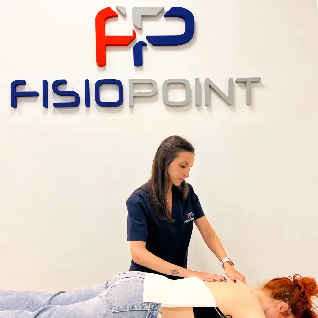
In some cases, when the problem is skeletal in nature, it is necessary to resort to surgery with an osteotomy to restore the correct joint relationships.
Book a Free Visit

Contact

Fill in the form,

You will be contacted as soon as possible


Physio point SErviCes

Physiomag

THERAPIES AND TREATMENTS

en_GBEnglish

*TO BOOK ONLINE, YOU MUST REGISTER WITH DOCTOLIB*

ALTERNATIVELY