What does the knee look like?
The knee is one of the most important structures in our body. It is the middle joint of the lower limb and supports the weight of the body during all movements throughout the day. Good knee alignment is essential, for example, when walking.
The joint is made up of four separate bones:
- Femur
- Tibia
- Perone
- Rotula
The fulcrum, however, lies in the joint of the femur and tibia. The articular surfaces of these two bones, in fact, fit together through two precise portions, namely:
- Condili femorali
- Piatto tibiale
Between them, two ring-shaped elements, called menisci, are interposed. The menisci are composed of fibrocartilage and their function is to improve the joint relationship of the two aforementioned bony structures and to absorb external shocks. They also reduce friction caused by the continuous movement of the joint heads. More specifically, the menisci are called:
These elements (both bones and menisci) are held together by numerous ligaments that create the structure of the joint capsule. The ligaments of the knee joint are:
- Lateral collateral
- Medial collateral
- Crociato anteriore
- Crociato posteriore
The first two are placed along the sides of the joint capsule and go to limit and protect the knee along the horizontal plane of motion. The two cruciate ligaments, on the other hand, prevent extreme hyperextension and hypereflexion movements of the knee, keeping it on axis in the frontal plane.
Also present in the knee are the joint bursae. They are actual sacs containing synovial fluid and have the purpose of further reducing the friction on the joint that can arise from external thrusts and movement forces generated by the constant contraction of the muscles. In the specific case of the knee, the joint bursae also have the function of reducing friction caused by the movement of the patella (or knee cap). The main bursae located along the knee joint are:
- Sovrapatellare
- Infrapatellare
- Prepatellare
What movements does the knee make?
The knee is a 'hinge' diarthrosis (a joint consisting of a cylindrical surface and a hollow) that performs two basic movements:
There are also very few degrees of lateral movement (not measurable) and rotation (approximately 45° total) of the tibia with respect to the femur, which provide the joint with three-dimensionality of movement in all axes and greater mobility of the leg.
Knee flexion
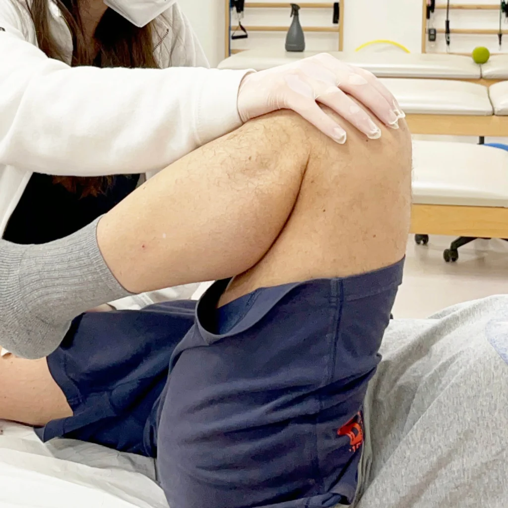
Flexion, in the knee, is defined as the movement that brings the back of the leg closer to the thigh. In a healthy knee, at the maximum flexion point, the tibia and femur form an angle of 140°. Such a wide range is possible due to the combined movement of rotation and sliding of the femoral condyles on the tibial plateau.
The muscles that enable flexion are located at the back of the thigh and are:
- Bicipite femorale
- Semitendinosus
- Semimembranoso
- Sartorio
- Gracile
- Popliteo
- Gastrocnemius
Knee extension
In contrast to flexion, extension consists of moving the posterior aspect of the leg away from the thigh and corresponds to grade 0. In particularly lax individuals, a hyperextension of up to approximately 10° can be appreciated. Extension is, similarly to flexion, possible due to the particular movement of the femoral condyles on the tibia.
The extensor muscles compensate for the flexor muscles and create balance with them. They are located at the front of the thigh and are:
- Retto femorale
- Vasto mediale
- Vasto intermedio
- Vasto laterale
All together, they are also known as the quadriceps femoris.
What are the typical problems of the knee joint?
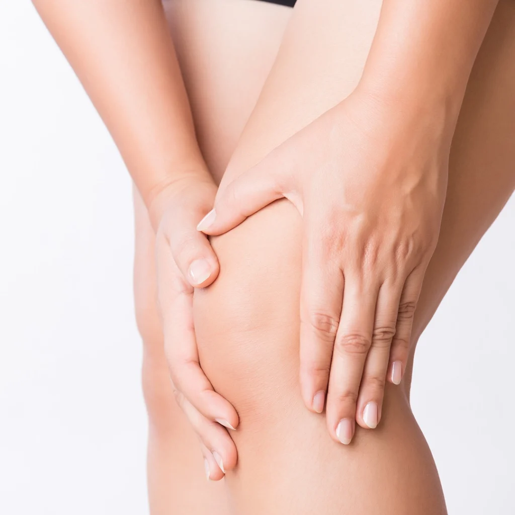
Since the knee, as we have already mentioned, is a joint that is extremely exposed to the stresses of gravity and external shocks, it very often runs into various problems. The first of these is, without a doubt, arthrosis. The knees have to constantly bear our weight, and because of this, over time, the cartilage that supports them degenerates, causing pain and difficulty in mobilisation. This phenomenon is called arthrosis.
There are also other problems, mainly of a traumatic nature. It is not uncommon, during sporting activity or even due to inattention, for some of the ligaments of the knee to suffer an injury or complete rupture.
Below, we look more specifically at some of the most common injuries.
Anterior cruciate ligament injury
The anterior cruciate ligament (ACL) keeps, together with the posterior cruciate, the tibia in axis with respect to the femur. More specifically, it limits movement anteriorly. It is certainly one of the ligaments most prone to injury and rupture. The causes can be many and in most cases occur during sporting activity. They are, consequently, very common in athletes. They include:
- Knee hyperextension
- Unnatural torsion of the tibia
- Direct trauma
Medial collateral ligament injury
The medial collateral ligament (MCL) keeps the knee joint stable and compact in the lateral-internal area. Under normal conditions, it prevents the knee from excessive valgus and lateral movement of the tibia. The causes are very similar to those we have defined for the cruciate ligament since, in this case too, the knee is brought into an extreme position so that the ligament cannot hold and ruptures. Collateral ligament injury occurs due to:
- Direct trauma
- Fall with fixed point on foot
- Excessive torsion of the tibia in valgus direction
Meniscal injury
The menisci in the knee act as 'shock absorbers' for the joint and allow smooth movement without hindrance or pain. Both menisci, however, can suffer wear and tear and injuries of varying severity due to:
- Cartilage degeneration
- Repeated incorrect movements
- Direct hits on the knee
In the context of meniscal injuries, we can identify different types of meniscal injuries that, depending on their severity, may lead to an acute knee lock. The most frequent injuries are:
- Horizontal
- Radials
- Longitudinals
- Flaps
- Bucket-handled
Conservative and surgical treatment of the knee
The choice of treatment between conservative and surgical depends, of course, on a multitude of factors. They are evaluated:
- Severity of injury
- Type of problem
- Patient's lifestyle
- Subject's profession and sporting activity
- Age
Physiotherapy for the knee
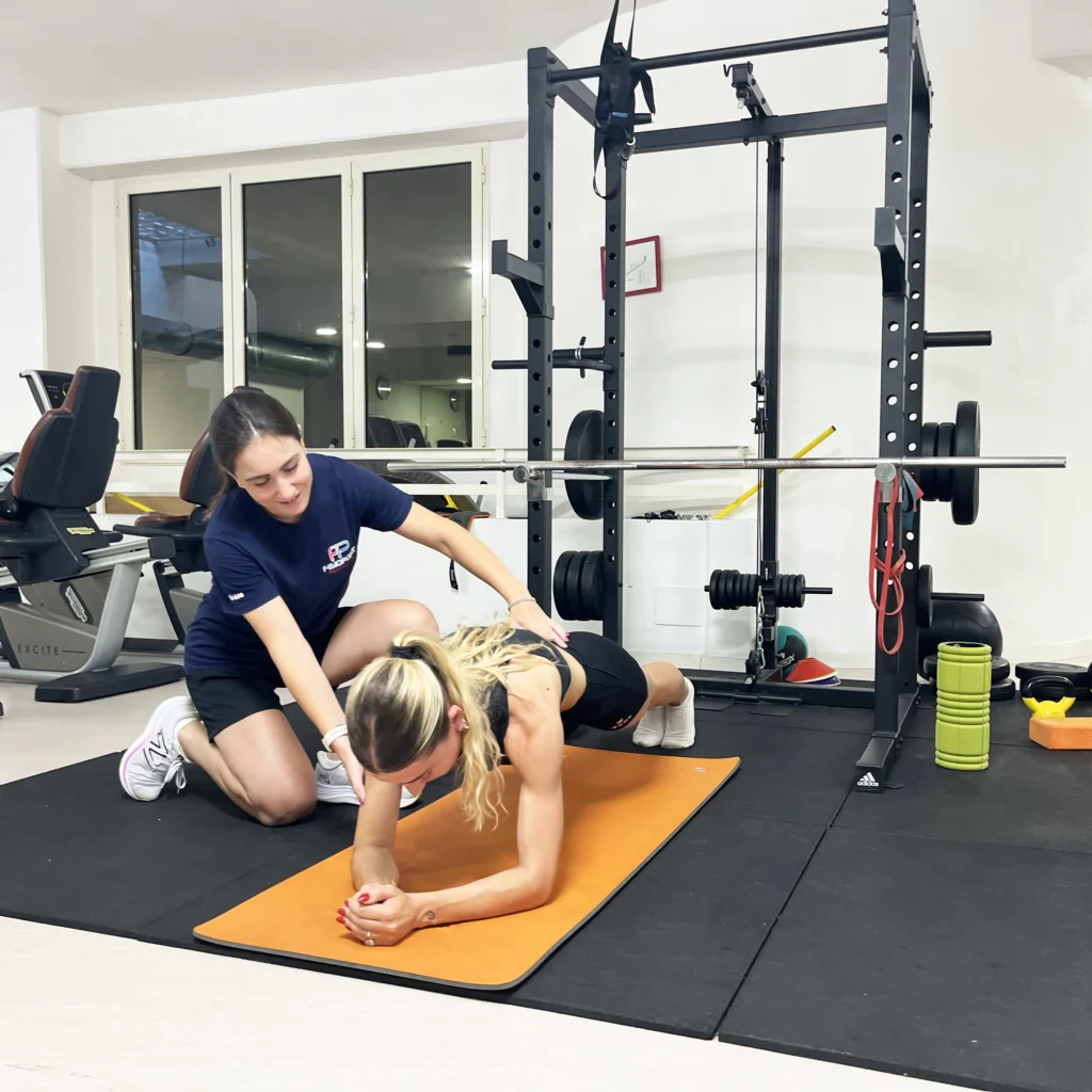
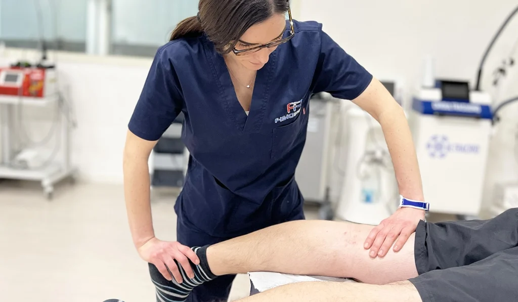
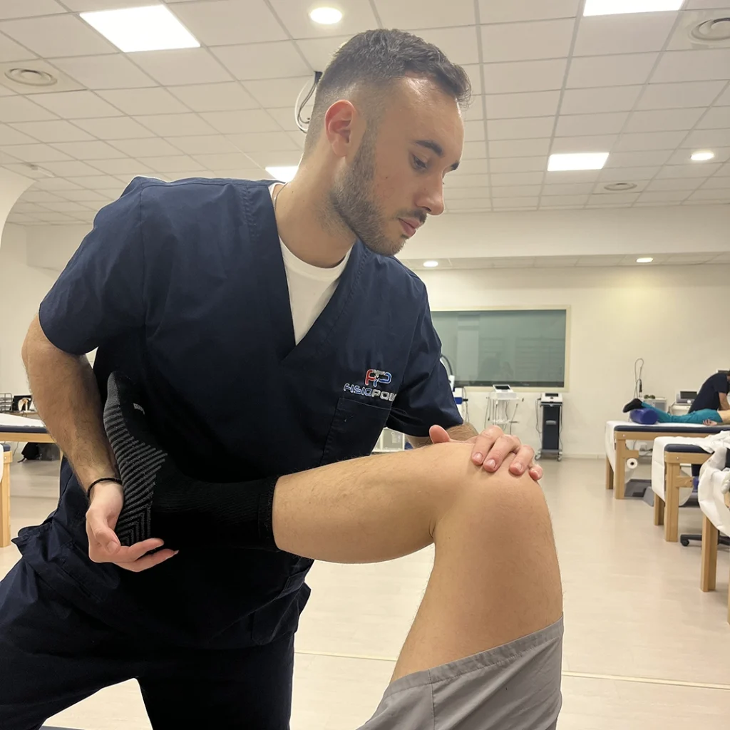
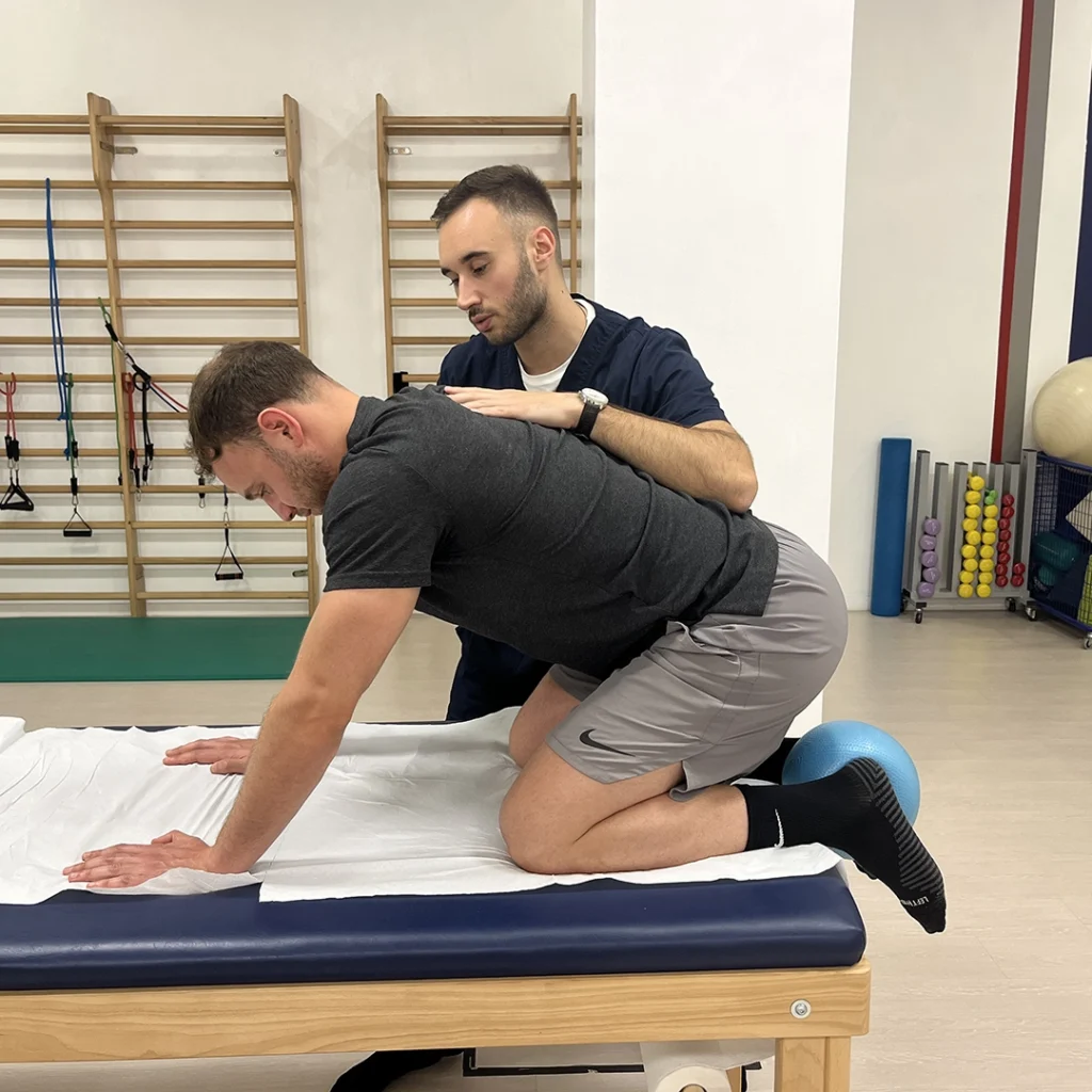
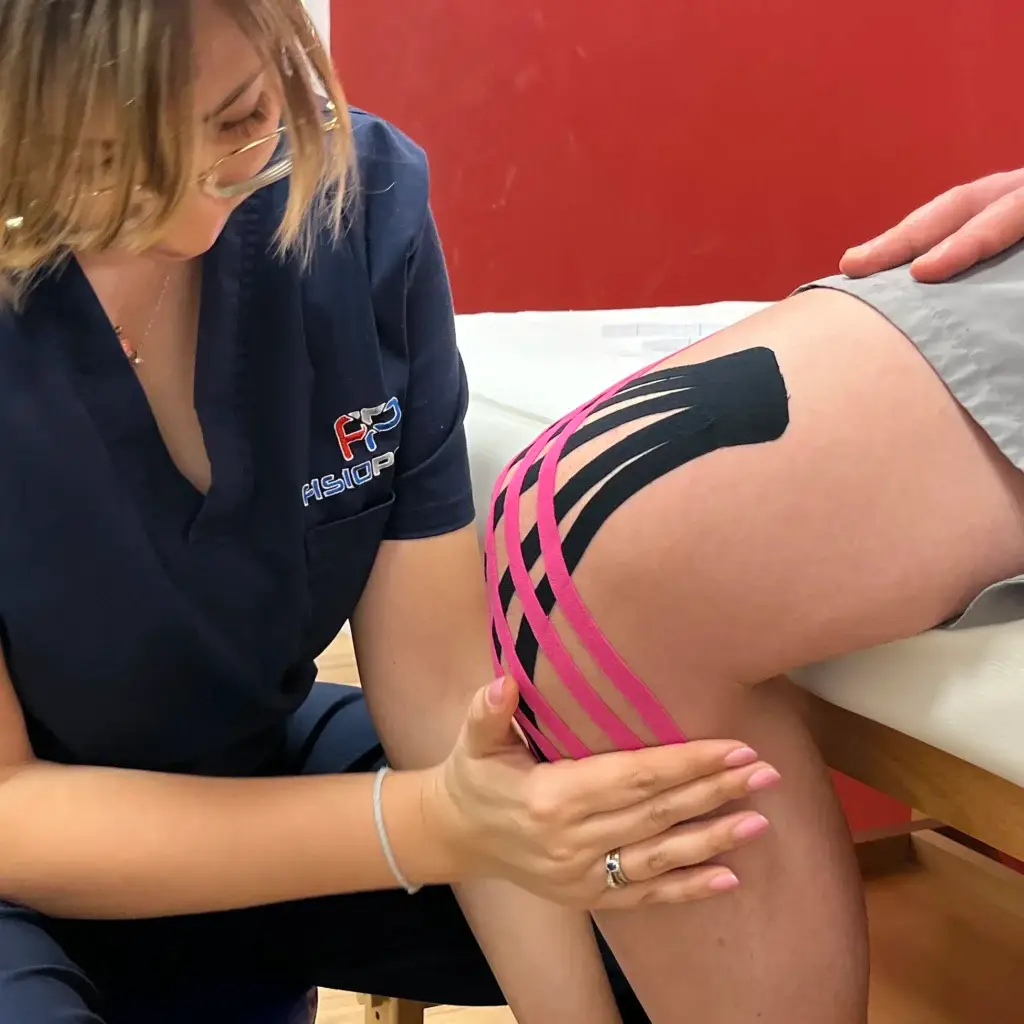
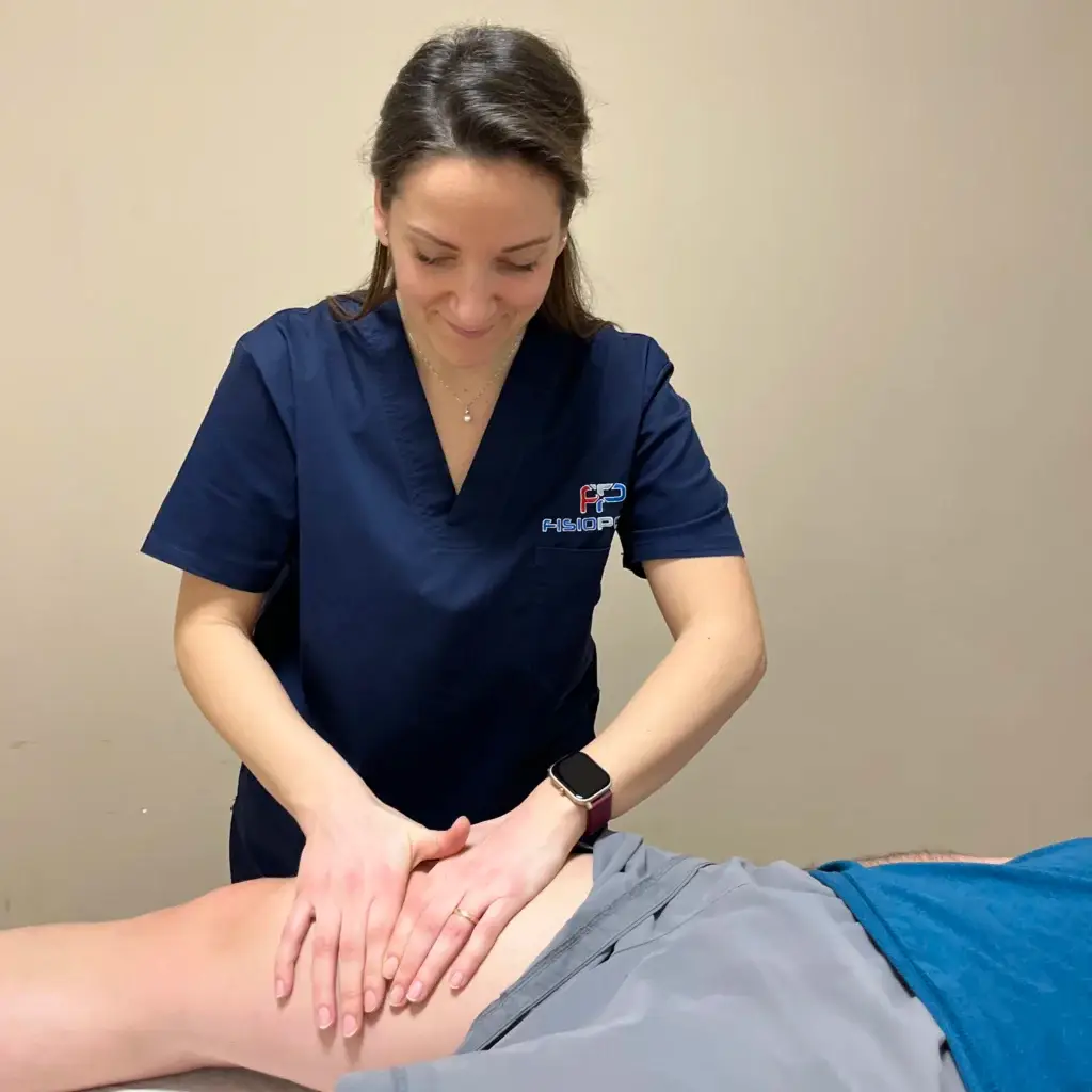
If conservative treatment is chosen, it will focus on:
- Pain control
- Drainage of swelling
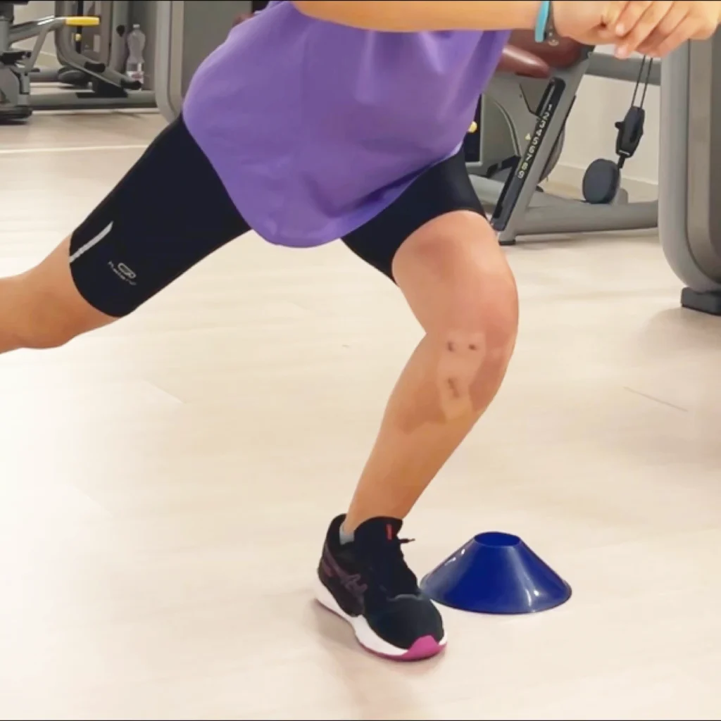
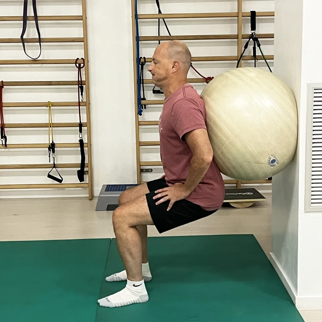
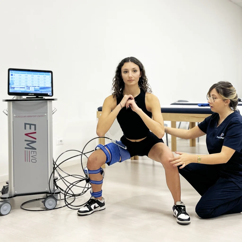
- Lower limb muscle strengthening
- Recovery of leg muscle tone
- Restoring joint mobility
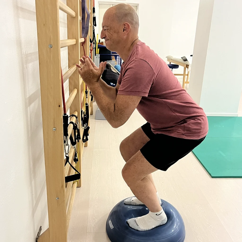
- Walking and proprioception re-education
- Return to physical activity and/or sports
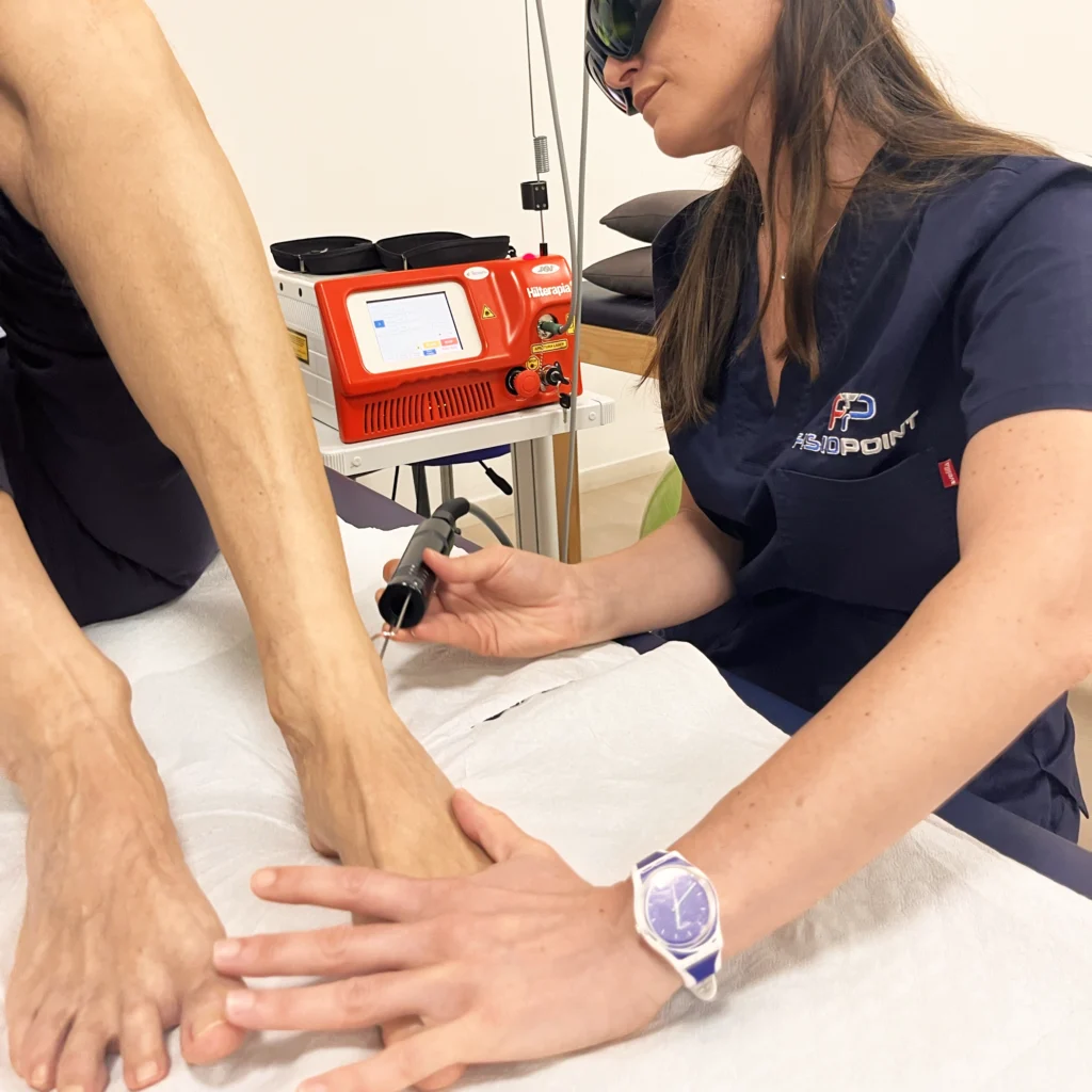
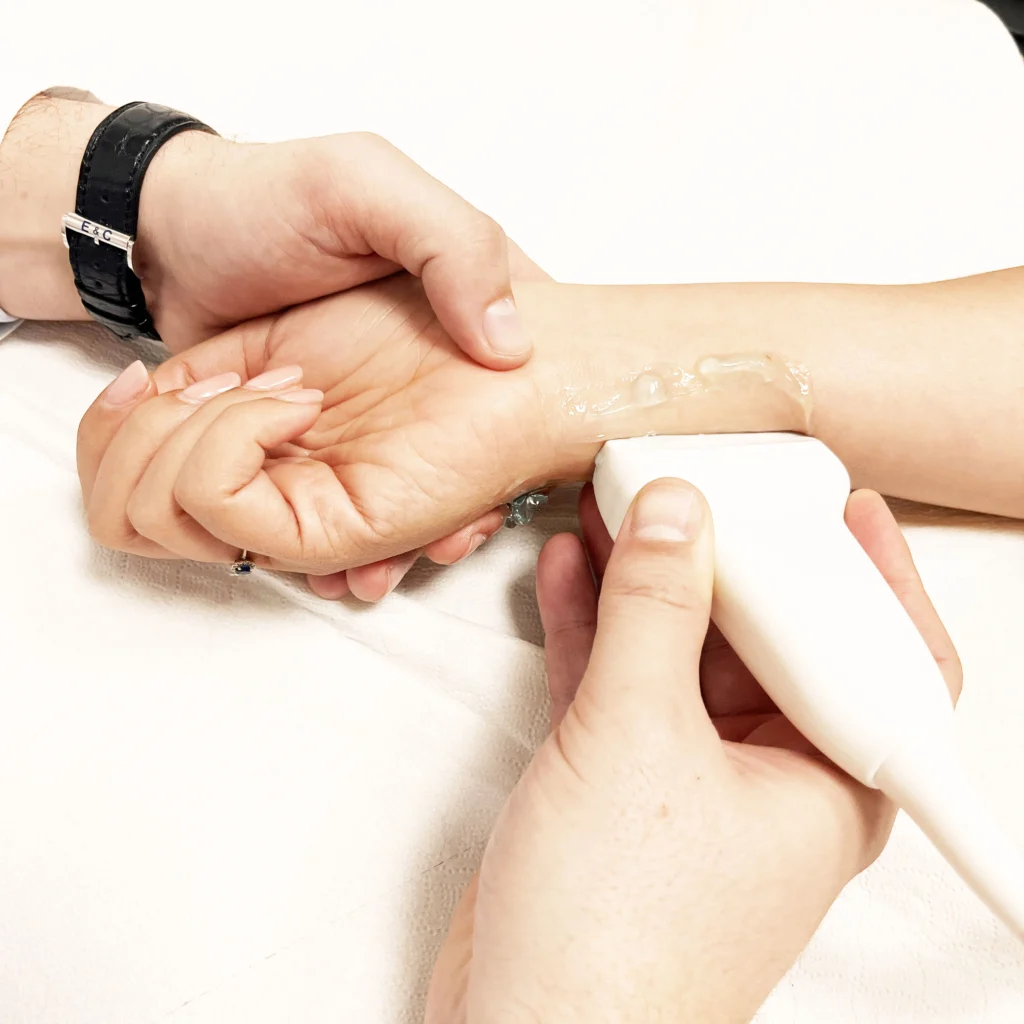
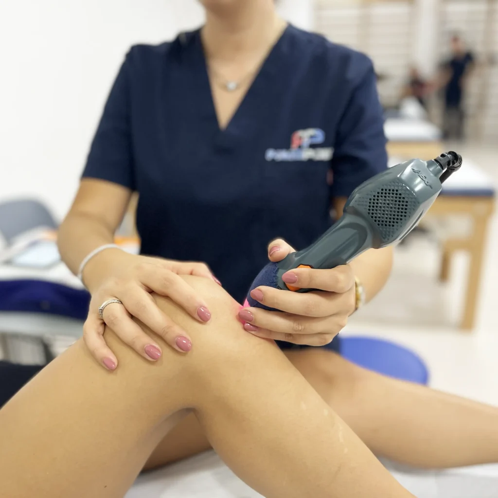
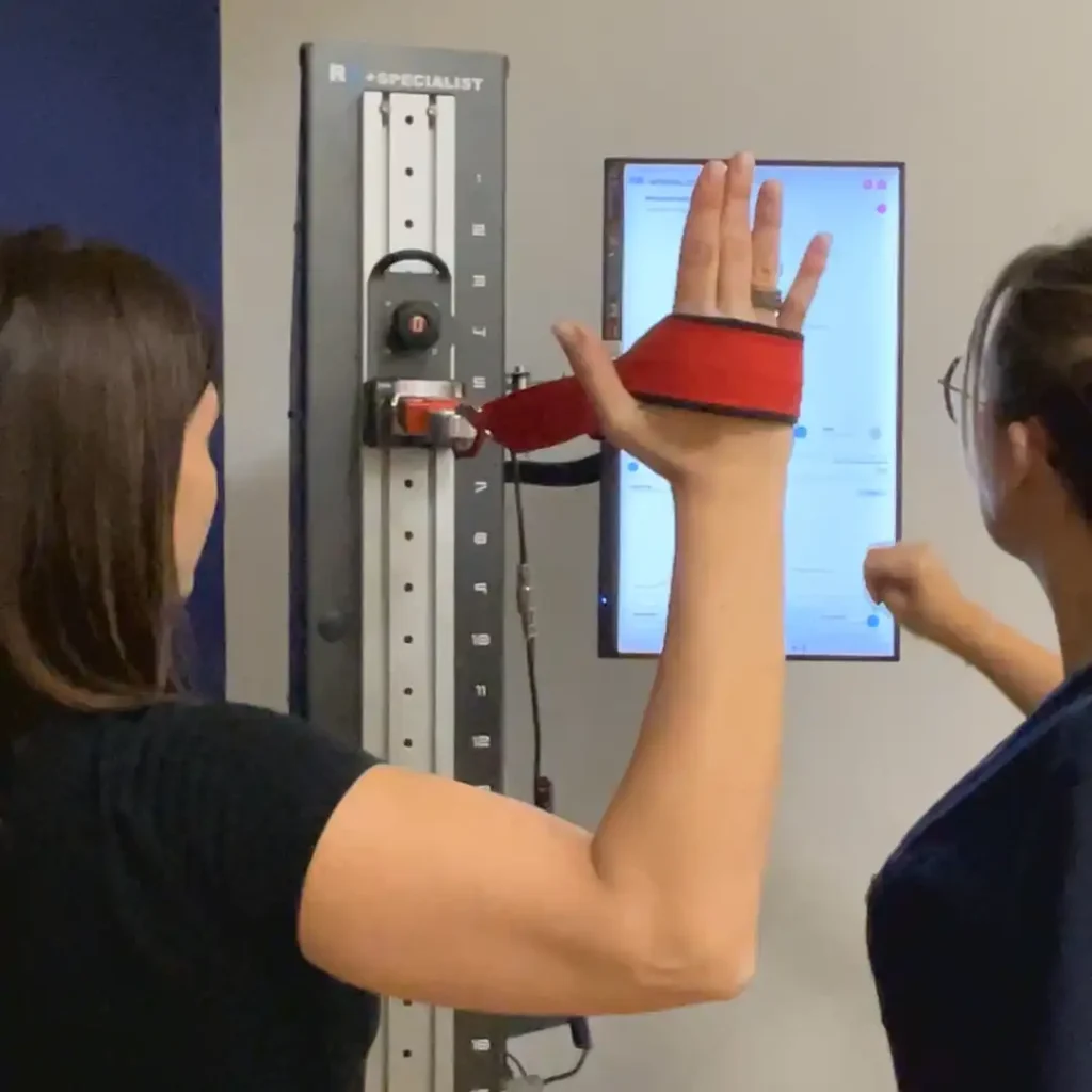
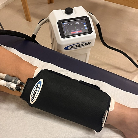
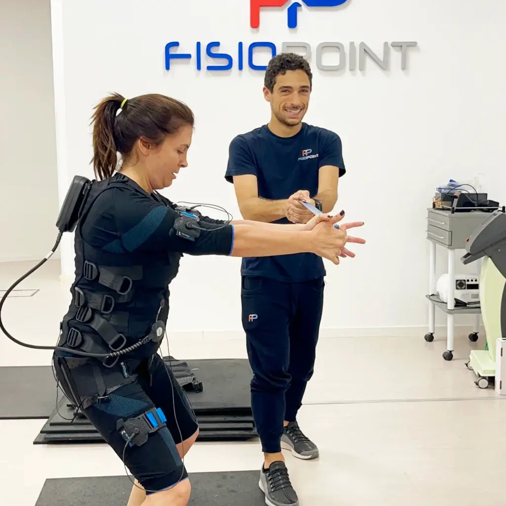
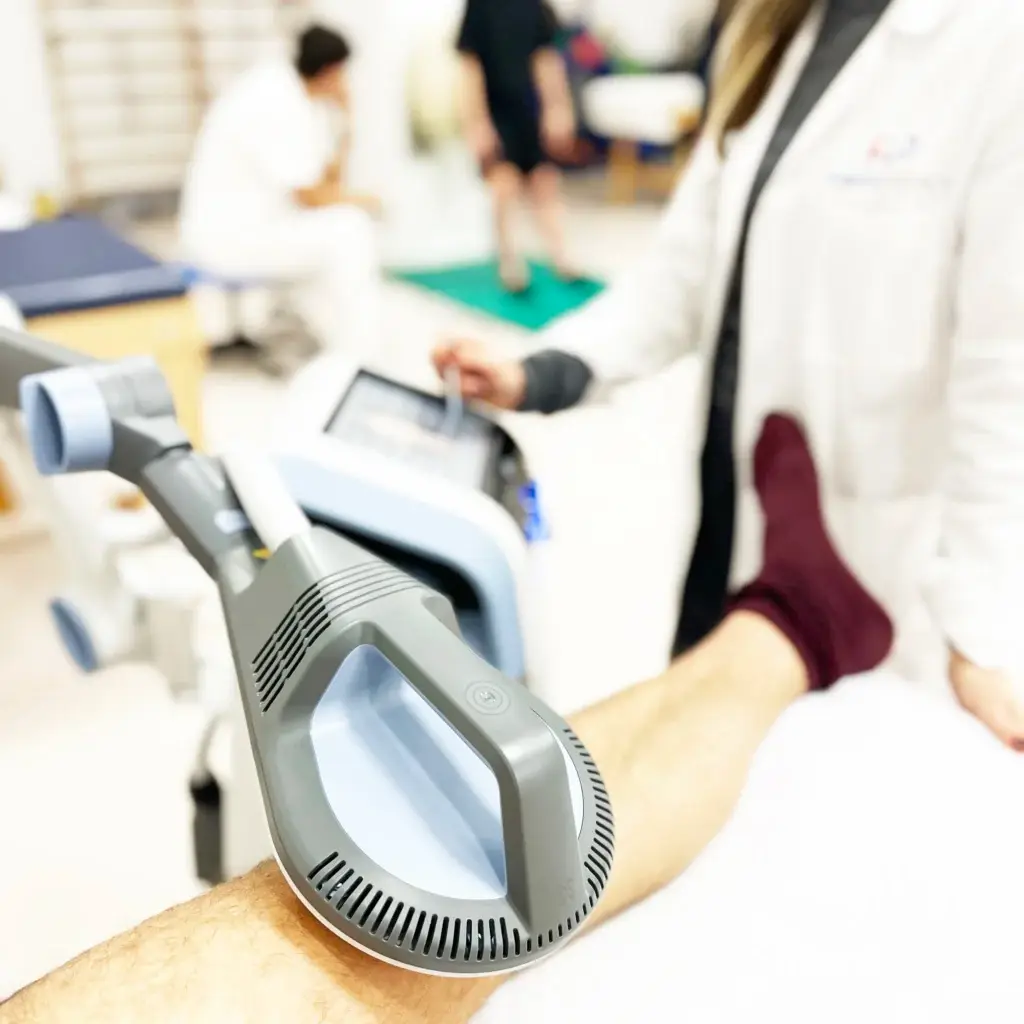
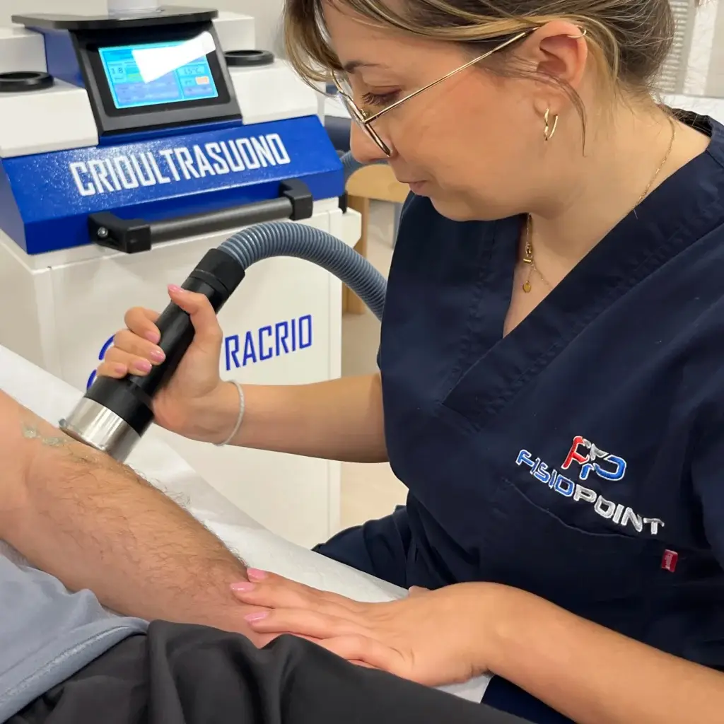
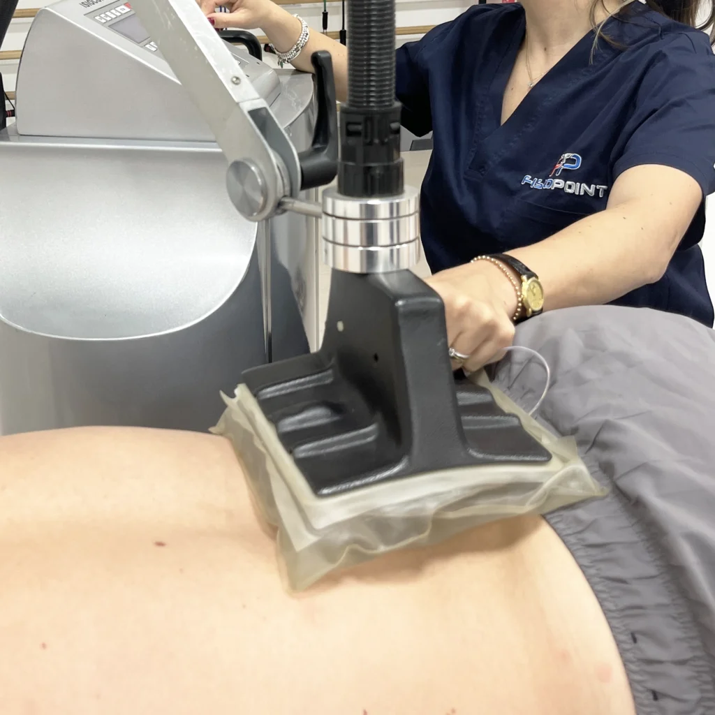
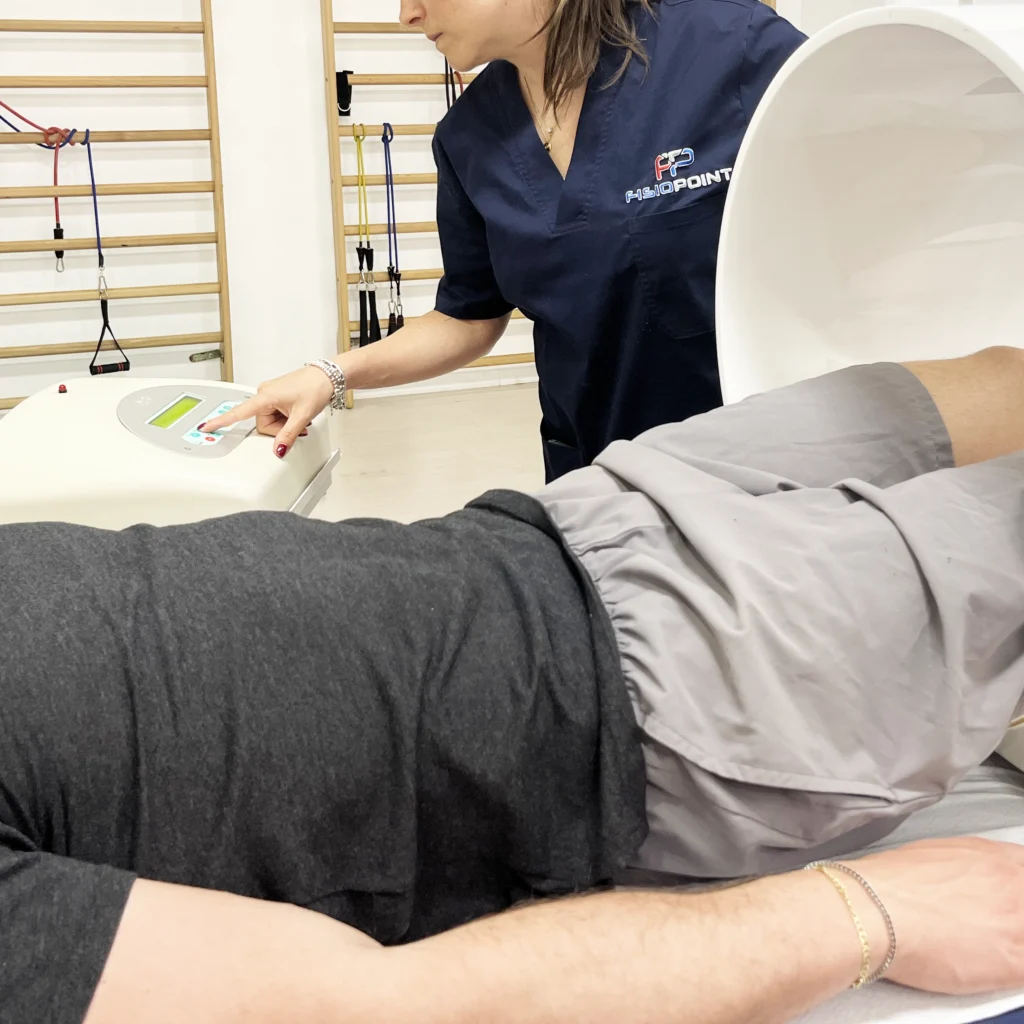
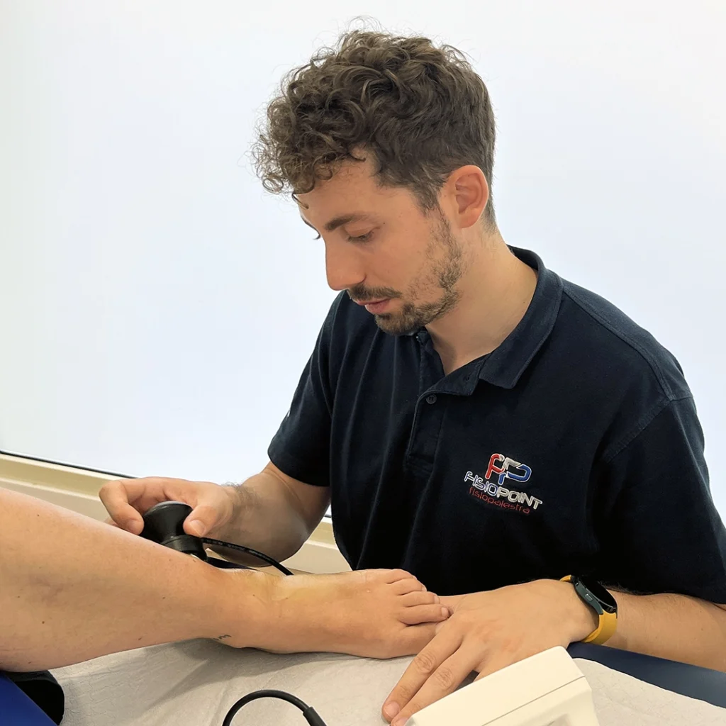
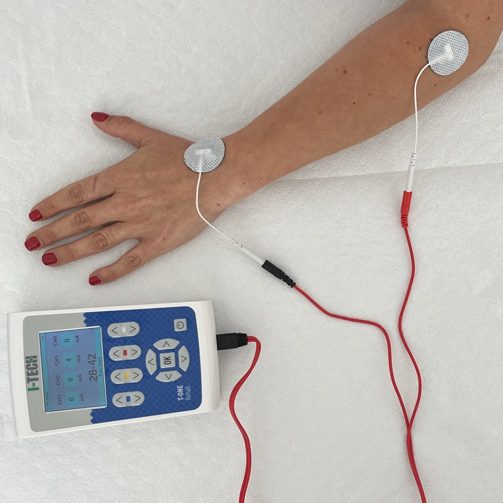
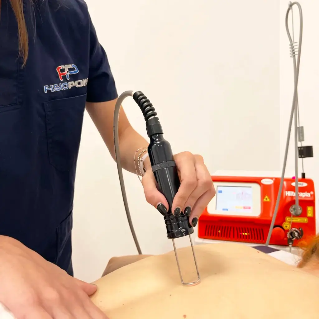
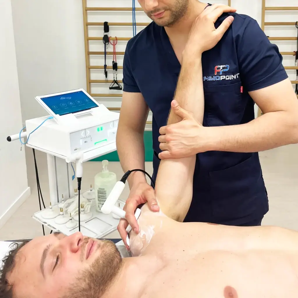
The aim of conservative treatment is to restore the joint to a normal state without the use of surgery. To do this, suitable exercises must be performed to strengthen all the muscles of the leg, which will provide the necessary support and stability. In the first phase of this treatment, it will be possible to speed up the healing process through the use of instrumental therapy such as:
- Tecar
- YAG laser
- Crioultrasuono
- Ultrasound
Knee surgery
In general, surgery is approached when the patient, following injury or accident, is very young and is engaged in sporting activity involving intense stress on the knee and frequent direct contact with it. Another category that always undergoes the operation is professional athletes, in order to regain optimum performance on the field with as little risk as possible.
In the case of injury to one or more ligaments, the surgeon reconstructs them by harvesting parts of the subject's muscle tendons, creating grafts from:
- Patellar tendon
- Semitendinosus
- Gracile
In cases of meniscal injury, on the other hand, one proceeds with the removal of one or more parts of the injured meniscus. This process is called meniscectomy.
Another very common operation, but one that is performed for different causes, is the knee replacement. It is implanted in mostly elderly individuals who have reached a high stage of arthrosis that makes their lives disabling.
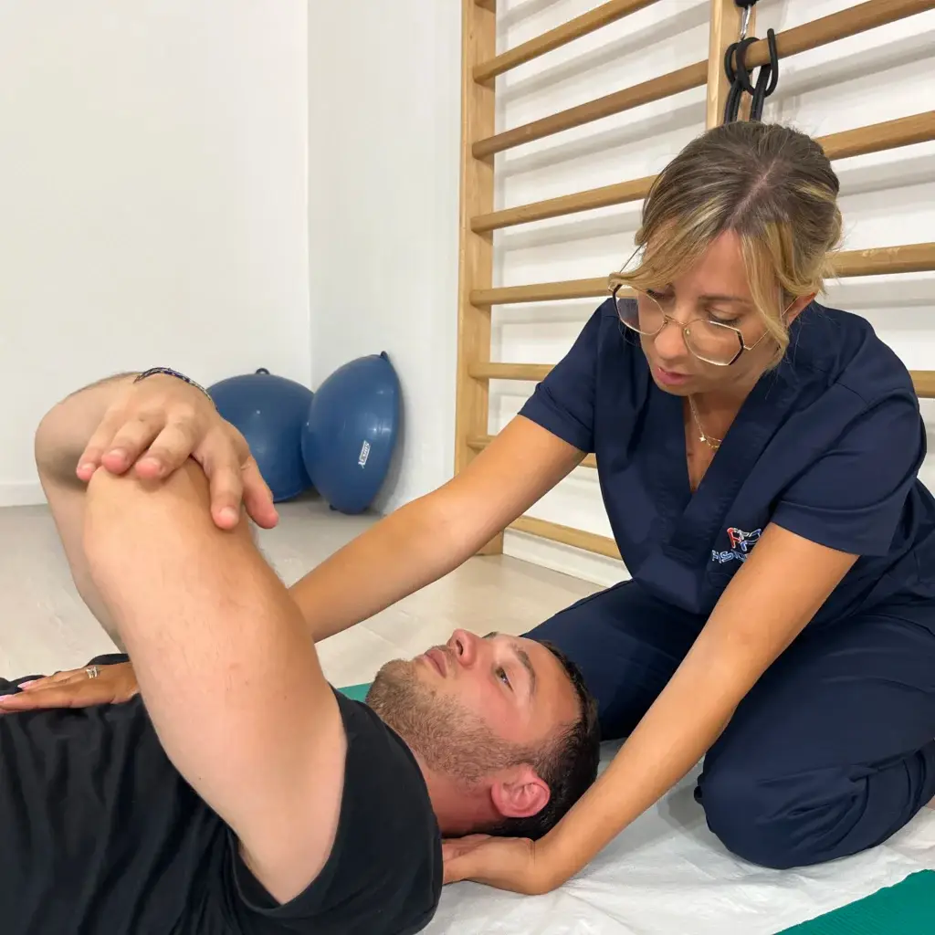
It is clear that, after any knee surgery, a course of physiotherapy rehabilitation is required, which will aim to completely restore movement and function, with a return to daily and sports activity in the absence of pain. The objectives and modalities are very similar to those of conservative treatment, but the course will be longer, as the implant and tissues must be given time to heal completely. The result, however, will be extremely better in all respects.
Text by Alessandra Burelli
Learn more by watching the videos on our channel YouTube






 English
 Italian