Anatomy of the knee and baker's cyst
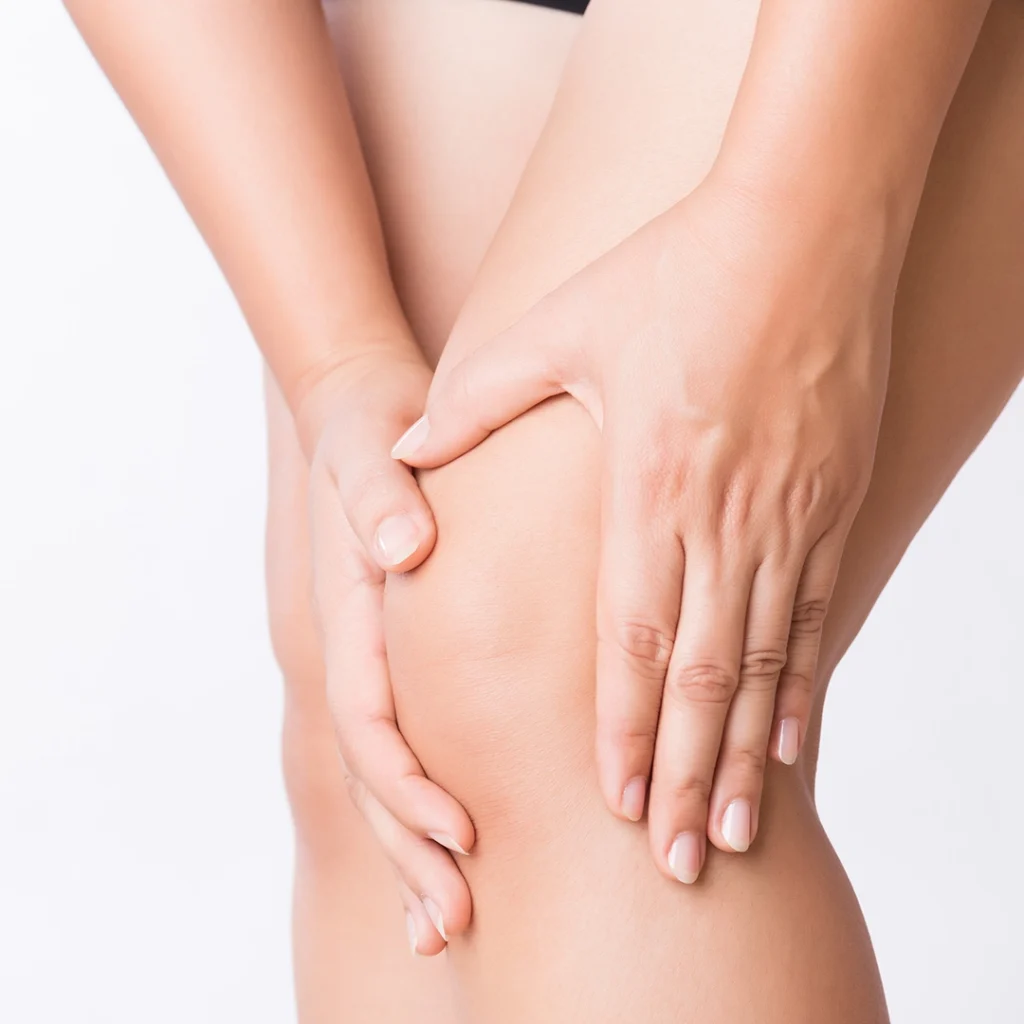
A Baker's cyst is an outward swelling filled with serous fluid. It generally appears at the back of the knee near the popliteal cavity. In fact, Baker's cyst also goes by the name 'popliteal cyst'.
It varies in size, ranging from a few millimetres to several centimetres, and can take the shape of a walnut, becoming clearly visible as well as palpable. It generally affects only one knee and is formed by an overflow of serum from the bursa poplitea.
Anatomy of the knee
In order to better understand what a Baker's cyst is, one must look at the anatomy of the knee.
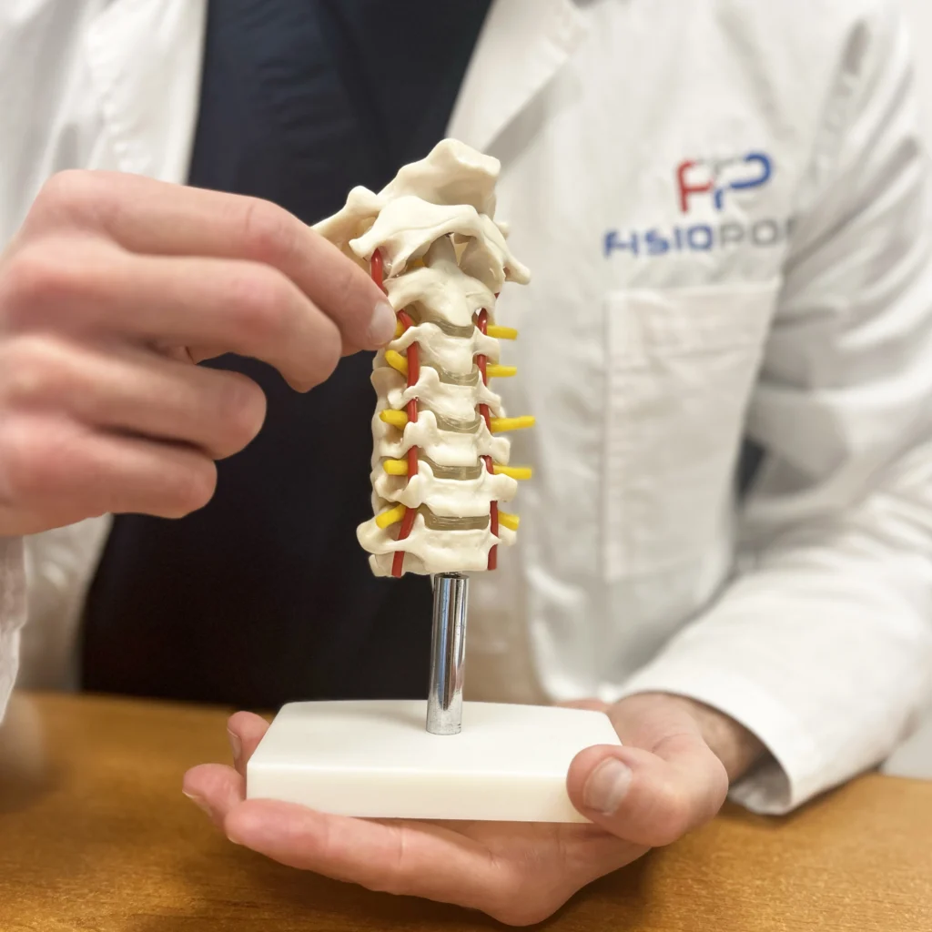
The knee is primarily formed by the articulation of two bony structures: the femur and tibia, but there is also a sesamoid bone, the patella, at the front. Interposed between the femur and tibia are the menisci: the inner meniscus and the outer meniscus, with the function of stability and absorption of stresses from the upper body. The joint is protected and encased by a capsule, which is lined on the inside by the so-called synovial membrane. The latter secretes and contains synovial fluid, which has a lubricating function, thus preventing rubbing between the bony structures during movement. The knee is also stabilised by ligaments, both inside and outside the joint, and muscles that provide active stability. Anterior and posterior to the knee, serous sacs are interposed between the ligamentous-muscle structures to reduce friction.
At the back of the knee is the popliteal bursa. The leakage of serous fluid from this bursa creates a sac, more external, in which this serum collects.
Causes and symptoms of Baker's cysts
There are two causes that lead to the formation of Baker's cysts, which can consequently be divided into two groups:
- idiopathic or primary Baker's cyst: it has no precise cause and is probably due to trauma or repeated stresses on the knee. Therefore, an abnormal accumulation of fluid in the back of the knee is produced. Children as young as 4-7 years old may suffer from it;
- secondary Baker's cyst: due to other pathologies such as meniscus problems, arthritis and arthrosis. In this case, there is excess fluid production, resulting in increased internal pressure and cyst formation. Adults between 40-70 years of age suffer from it.
Such a formation is benign and obviously the picture of symptoms varies from person to person. Certainly, the resulting discomfort is related to the size of the cyst. In fact, when it is small, it may even be asymptomatic. In more severe cases, on the other hand, it can occur:
- a pain in the knee that reaches the calf
- joint stiffness with difficulty in performing normal everyday movements
- a swelling that extends to the entire joint area.
Rarely, a rupture of the cyst may occur, resulting in fluid escaping to the nearby muscles and obvious reddening and swelling of the entire area. In other cases, the cyst may calcify or be a disturbing factor for the nerve structures that pass through the back of the knee.
Diagnosis of Baker's cysts
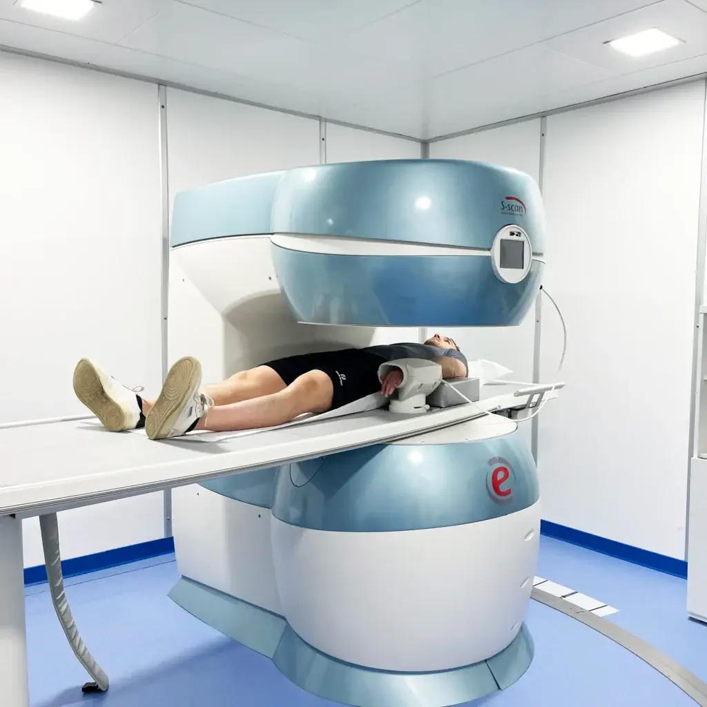
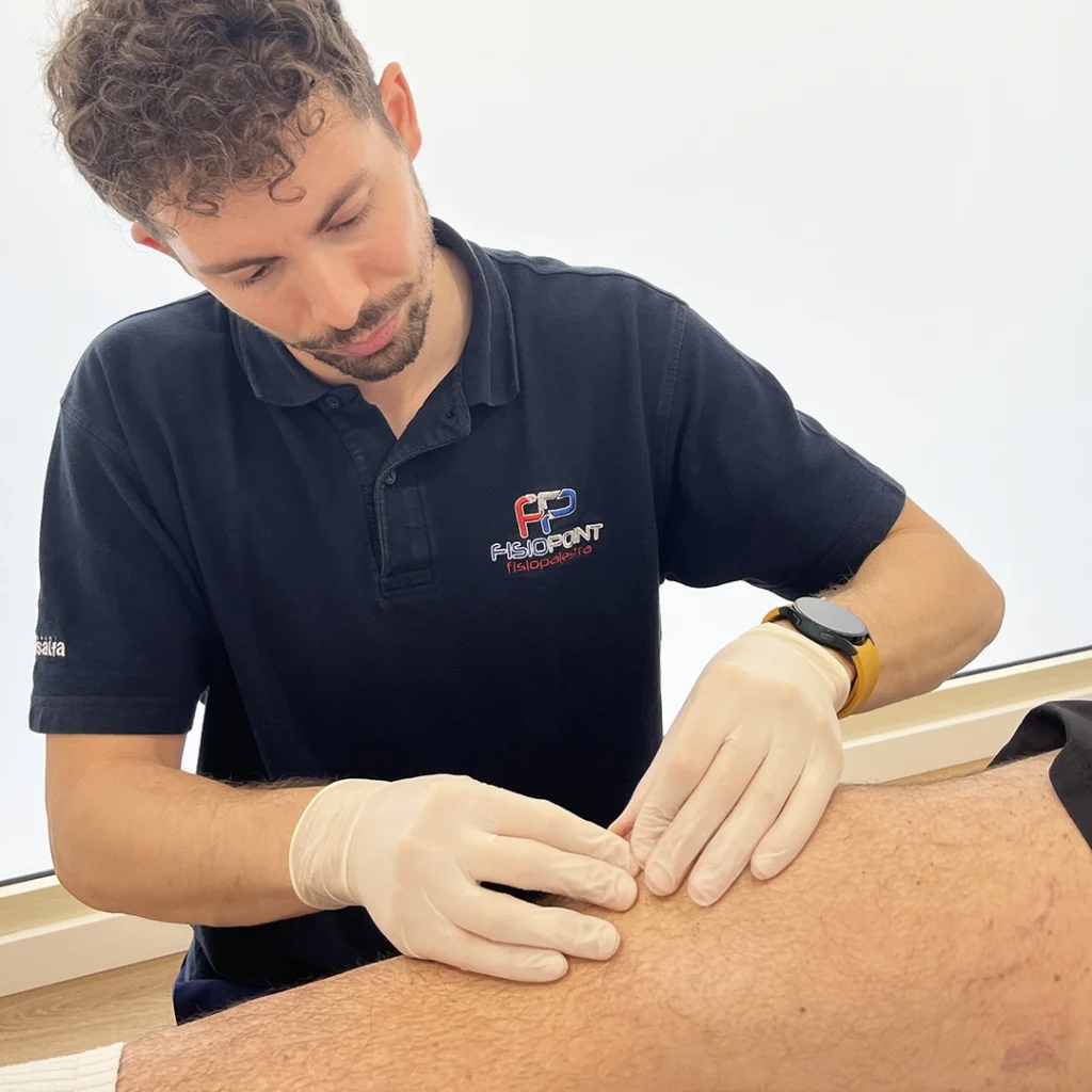
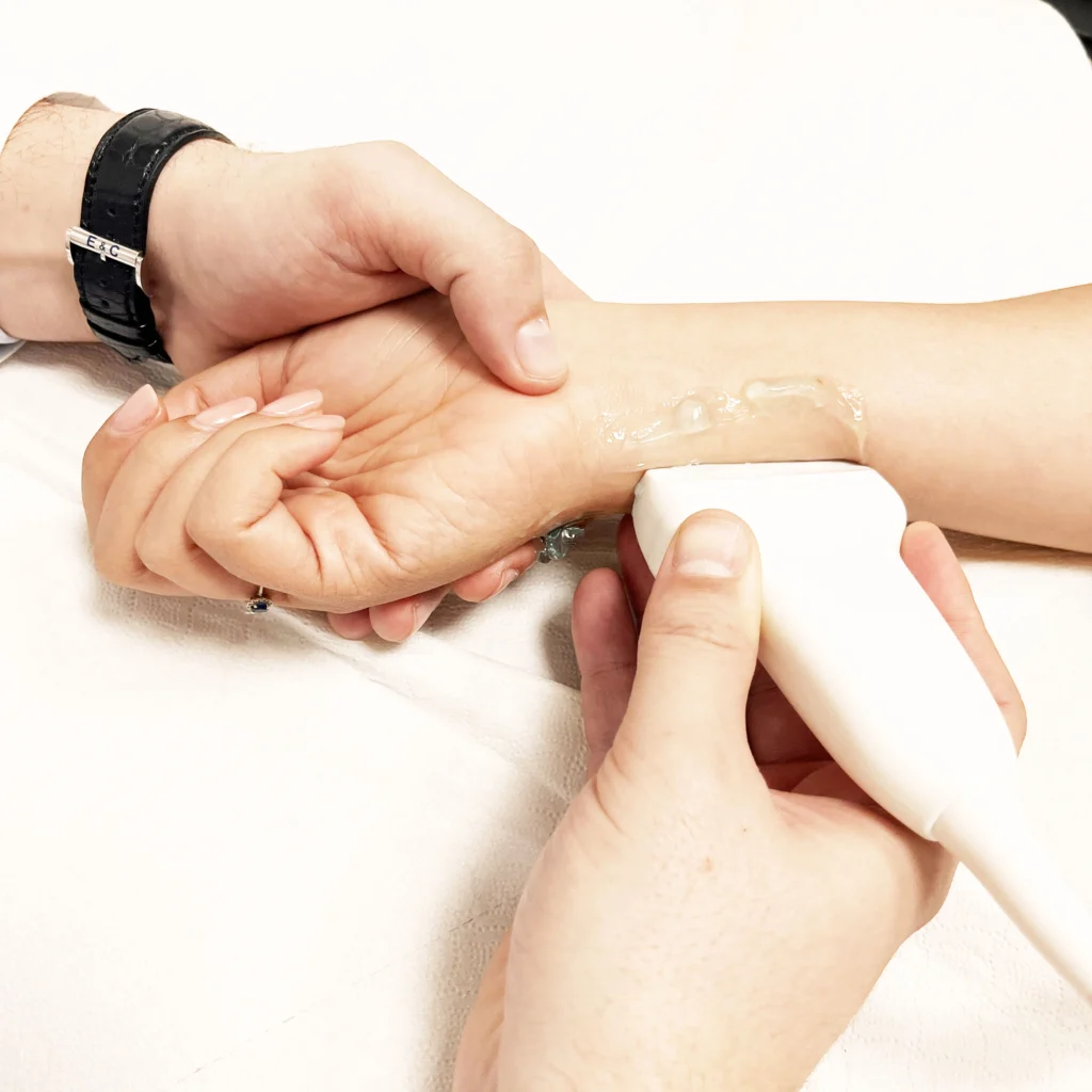
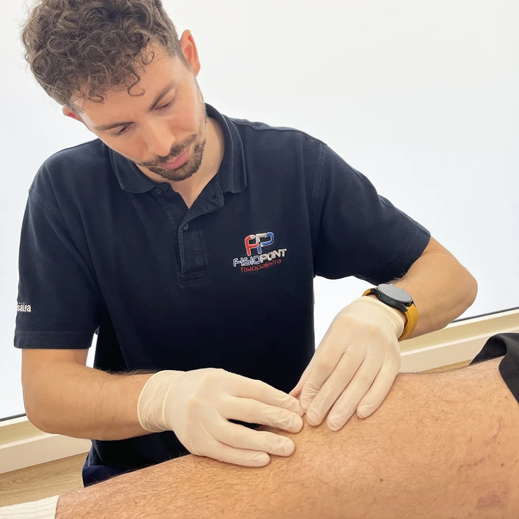
The cyst is visible to the naked eye or palpable. Even, in some cases, with reflected light it is possible to glimpse the liquid contained inside. If such a cyst occurs, the best thing to do is to consult a doctor or a good physiotherapy centre. The specialist will then direct the patient to a more thorough examination to rule out other pathologies. There are two instrumental examinations that can be performed:
Treatment and care of Baker's cysts
Treatment is not always required. In young people, it is possible for the cyst to reabsorb on its own and such patients have a healthy knee.
The problem arises in adults, where the knee is no longer structurally intact and there is an overproduction of fluid, due to chronic joint inflammation or a ruptured ligament or meniscus. In this case, there are several ways to intervene:
- aspirate the fluid present in the cyst;
- inject cortisone-based substances to try to reduce inflammation;
- intervene on cartilage or ligament problems, if it is thought that these may be at the root of the problem.
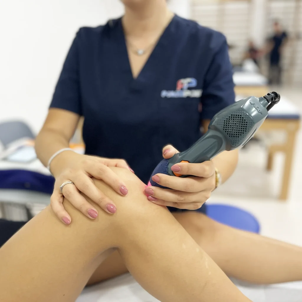
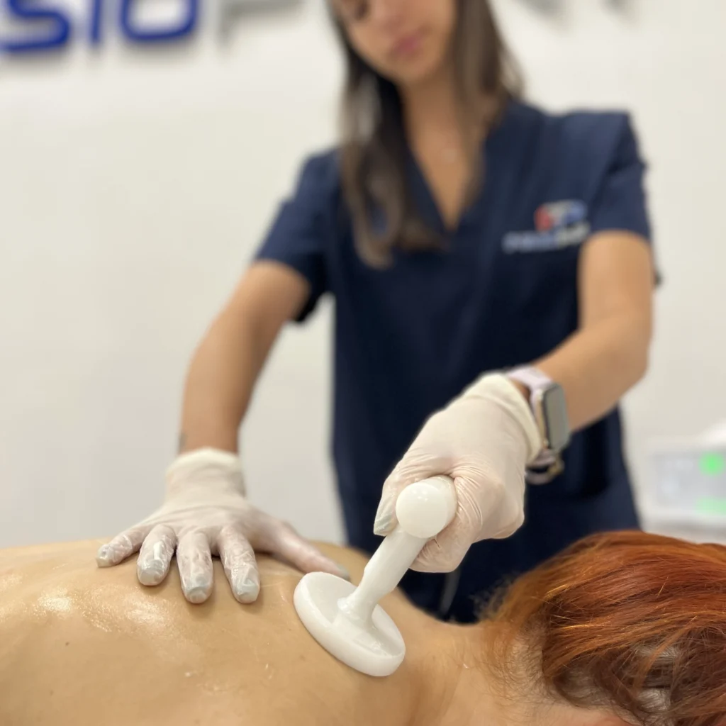
In some patients, where acute pain is present for long periods, it is possible to remove the cyst surgically using more or less invasive methods. In most cases, it is possible to go to a good physiotherapy centre and start a rehabilitation course. In fact, there are machines that can reduce inflammation and increase fluid drainage:
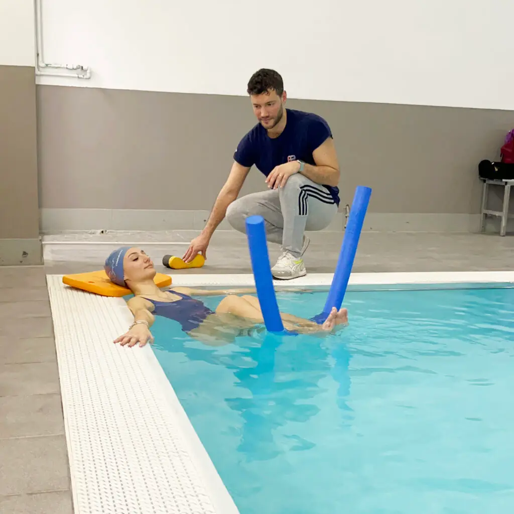
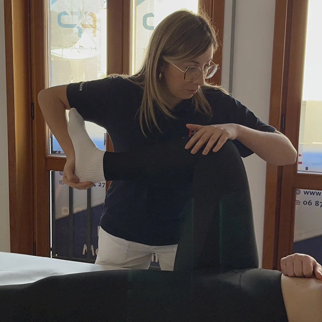
In a second step, one can then work on the mobility of the joint in a cautious manner and try to re-establish normal range of motion.
Exercises for treating Baker's cysts
Finally, the third phase involves strengthening the muscles of the knee, without increasing the loads and stresses. Below are some examples of exercises:
- From a supine decubitus position (lying belly upwards), while the healthy leg is bent with the sole of the foot resting on the ground, the other leg performs some sprints (with the leg outstretched and the hammer foot), bringing the thigh up to the height of the contralateral knee;
- The same exercise just described can be performed with the leg extended and locked at the level of the contralateral knee. The position must be held, in isometric contraction, for a few seconds;
- From the same supine decubitus position, this time with both legs bent, you can place a pillow or sponge ball between your knees and perform contractions by squeezing your legs together;
- the same exercise as described above can be carried out, maintaining an isometric contraction for a few seconds;
- in a lateral decubitus position, on your side, you can bend both legs (with the healthy leg resting on the ground), place both legs inside a looped elastic band and perform upward limb openings or hold the position, in openness, for a few seconds.
These are some simple strengthening exercises that can be performed safely.
It is important to always rely on qualified people.
Learn more by watching the videos on our channel YouTube


 English
 Italian