Femur fracture is a disabling and potentially dangerous event, especially in elderly individuals, due to its possible related medical complications.
The incidence is higher in women than in men and the treatment, unless there are special contraindications, is surgical and varies depending on the type of fracture. The type of surgery performed, the patient's age and clinical condition will determine the recovery time and post-operative instructions to be followed.
Hip: anatomical outlines
The femur is the longest and strongest bone in the entire human skeletal system. It provides insertion to several muscles essential for walking and, articulating proximally with the acetabulum (itself formed by the three pelvic bones ilium ischium and pubis), forms the hip joint, while its distal portion articulates with the patella and tibia to form the knee.
Looking at the articulation of thehipwe can see how this represents the point of union between the pelvis and the lower limb. This synovial joint is composed of the union of the head of the femur (which represents the proximal end of the latter bone), the acetabulum (structure of the pelvis) and a joint capsule with its synovial membrane, ligaments, two synovial bursae and several muscles that fit into it.
The function of thehip, and of the femur in general, is to support the weight of the upper part of the body and at the same time allow walking thanks to the movements that the lower limb can make.
The hip, also known as the 'coxofemoral joint', is considered a spherical diarthrosis, endowed with great mobility, which is characterised by a perfect fit between the concave surface of the acetabulum and the convex surface of the femoral head.
Anatomically, this structure is located anterolaterally to the gluteal region, below the iliac crest and above the great trochanter of the femur.
The hip, like all synovial joints, has articular surfaces with a coating of hyaline cartilage, which serves to facilitate the sliding of the two structures, prevent friction between them and cushion load forces.
What does the femoral head look like?
The femoral head, resting on what is known as the 'femoral neck', is a rounded, almost spherical bony protuberance that is located on the distal end of the femur and projects medially relative to it.
The head of the femur is covered with hyaline cartilage, with the exception of the central part, where there is a small rough depression known as the fovea capitis; the fovea capitis houses a very important ligament of the hip: the round ligament of the femoral head.
As mentioned earlier, the head of the femur inserts into the acetabulum, a structure also known as the 'cup', located in the outer portion of the ilium (formed in turn by the ilium, ischium and pubis.
The acetabulum consists of several parts:
- The acetabular labrum, a fibrocartilaginous structure that peritrally surrounds the acetabulum itself. It ensures stability and correct positioning of the femoral head and the hip as a whole
- The semi-lunar face of the acetabulum, covered with hyaline cartilage, is the only portion of the acetabulum that actually articulates with the femoral head
- The acetabular incisura, the lower portion of the surface that constitutes the acetabular notch, ensures the passage of the transverse acetabular ligament, in addition to giving insertion to the aforementioned round ligament, and forms the so-called acetabular foramen, necessary for the passage of the blood vessels that will supply the femoral head
- The acetabular fossa, the deepest and most central portion, is in continuity with the acetabular incisura and contains adipose tissue covered by synovial membrane
Hip: the joint capsule and ligaments
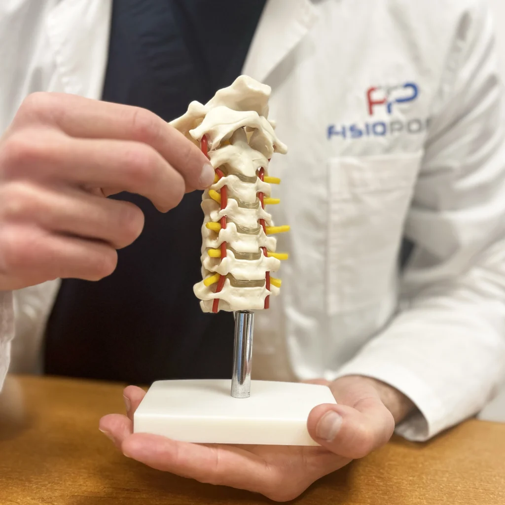
Like all synovial joints, the hip also possesses a joint capsule, which is a dense fibrous connective tissue structure that surrounds and holds together the two joint surfaces (in this case the acetabulum and the femur up to the neck).
Specifically, the joint capsule at the acetabular level inserts on the margin of the acetabular labrum and the transverse acetabular ligament, while at the femoral level it inserts:
- At the intertrochanteric line, anteriorly
- At the base of the femoral neck, above
- Approximately 1 cm higher than the intertrochanteric crest, posteriorly
- Near the small trochanter, inferiorly
The anterior and superior hip joint capsule is very thick and strong, due to the greater stress the joint is subjected to in this area, while its posterior and inferior portion is thinner and less strong (in fact, joint stress is lower in this area).
The (extracapsular) ligaments that stabilise and strengthen the hip structure are:
- ileofemoral
- ischiofemoral
- pubofemoral
The joint capsule is connected to these ligaments through connective fibres and, thanks to its synovial membrane, produces synovial fluid, which is essential for the lubrication of the various joint components.
The intracapsular ligaments, on the other hand, are located within the hip joint capsule and are:
- The roundness of the femoral head
- The transverse acetabular
Their function is to stabilise the joint, provide passage and protection for nerves and blood vessels that will innervate and irrigate the hip itself.
Specifically, the round ligament of the femoral head, which runs from the fovea capitis of the femoral head to the acetabular incisura (located on the acetabulum), in addition to providing stability to the hip joint, carries within it a branch of the obturator artery (also called the 'femoral head artery'). The purpose of this artery is to irrigate the femoral head and thus ensure its nourishment and survival.
The transverse acetabular ligament, on the other hand, is a flat, strong ligament that is located at the level of the acetabular incisura and here forms a bridge under which the 'acetabular foramen' is located, through which nerves and blood vessels destined for the hip itself enter the joint.
Synovial bursae
The hip has two synovial sacs:
- Synovial bursa of the iliopsoas
- Synovial bursa of the greater trochanter
The former is located between the joint capsule and the iliopsoas muscle, while the latter is located, as its name suggests, above the great trochanter of the femur
The innervation of the hip
The hip joint is innervated by nerves derived from the lumbosacral plexus (second lumbar to first sacral vertebrae), these nerves are:
- The femoral, which innervates the anterior portion
- The obturator, which innervates the lower portion
- The superior gluteal nerve, which innervates the upper
- The nerve of the quadratus femoris and inferior twin muscles that innervate the posterior portion
Precisely because of this sharing of innervating structures, a pain in the lumbosacral spine may radiate to the hip, while a hip-specific pain may also be felt at the knee level.
What are the hip muscles?
The muscles associated with the hip joint are:
- The buttocks (great, middle and small gluteus), located in the postero-lateral region of the hip
- The adductors (long, short, great and small adductors, external obturator, pectineus and gracilis), located in the inner hip region (inguinal area)
- The iliopsoas, which inserts on the small trochanter of the femur
- The rectus femoris and sartorius, located in the anterior region of the thigh
- The ischiocrural muscles (also called 'hamstrings'), i.e. the biceps femoris, semitendinosus and semimembranosus, located in the posterior region of the thigh
- The piriformis, external obturator, internal obturator, quadratus femoris, superior twin and inferior twin muscles, which together form the group of extrarotator muscles (external rotators) of the hip.
What is the hip joint used for?
The hip joint has two main functions:
- Supporting the weight of the body, both statically and dynamically (e.g. when walking), and discharging forces along the femur to the foot
- Allowing movements of the lower limb through activation and support of the above-mentioned muscles in the different directions (flexion, extension, intra and extra rotation)
What are the most common hip disorders?
The hip can be involved in numerous diseases and clinical conditions, some more common than others, such as
In this article, we will look at femoral head fracture and describe its general characteristics along with its treatment.
Return to normality and prevention
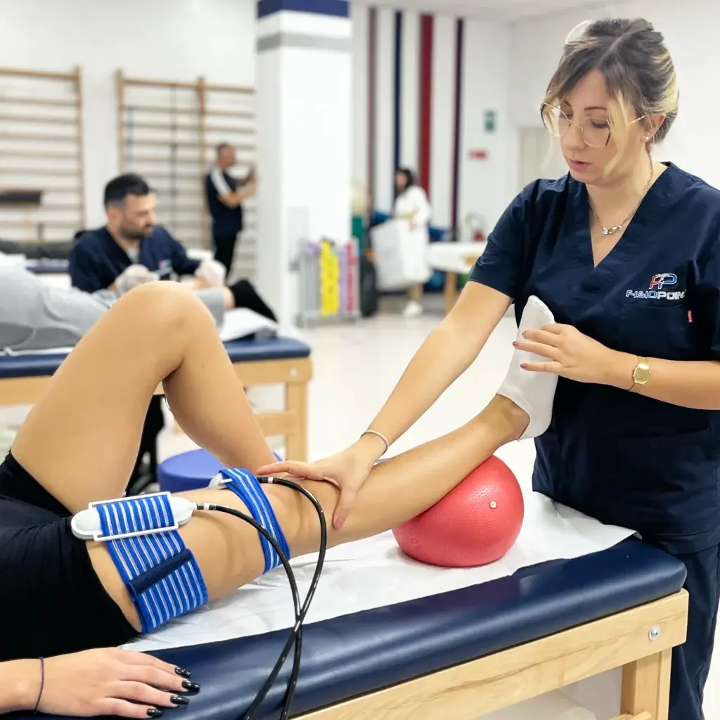
Femur fracture, both in young and old subjects, can bring with it physical and emotional consequences: in fact, after the fall, the patient may feel insecure in walking and the fear of falling again and dislocating the implanted prosthesis may lead him to move less and less and isolate himself from social activities. After surgery, it will therefore be essential for the patient to begin a rehabilitation programme to restore joint function, strengthen muscles, reset the gait pattern correctly, and improve balance and confidence in movement.
Particularly in elderly patients, it is important to keep a medical eye on possible complications such as thrombosis, phlebitis, infections, cardiac and respiratory problems, which can occur even after hospitalisation.
To prevent hip fracture, it is recommended to eat a diet rich in calcium and vitamin D to maintain bone health and to exercise regularly to maintain balance and muscle tone. To reduce risk factors, it is necessary to limit cigarette smoking and alcohol intake (both have negative effects on circulation and tissue oxygenation). It is also important, especially for older people, to reduce the risk of falls, e.g. by eliminating potential hazards and obstacles in the home that could cause them to trip or slip.
Text by Alessandra Del Vecchio
Learn more by watching the videos on our channel YouTube







 English
 Italian