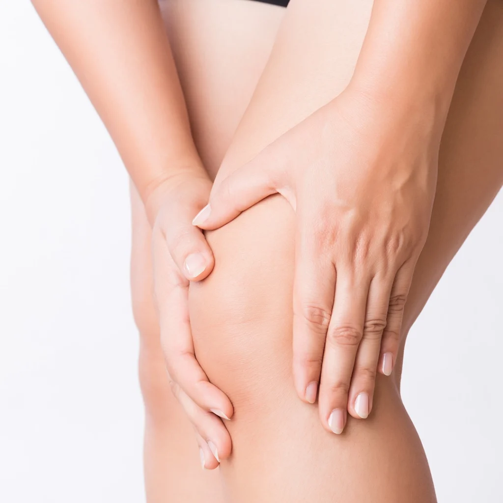
It often happens that one or both knees are in severe pain, severely limiting one's daily activities. When this pain is related to wear and tear of the segments that make up the knee, the joint is damaged and the possibility of movement is minimal, it may be advantageous to resort to the implantation of a knee prosthesis. The aim of a knee prosthesis is to decrease pain and increase articularity by replacing damaged skeletal sites with an artificial joint. It is a real operation that requires good preparation in the period leading up to it, and it is essential to have physiotherapy in the post-operative period in order to best achieve the goals set and to return to leading a normal life. There are two types of knee replacement: the total knee replacement, which is used most frequently, and the partial knee replacement. It is the surgeon who will perform the operation who chooses the type of prosthesis to be used, especially depending on the age and state of health of the patient.
The knee: outlines of anatomy and biomechanics
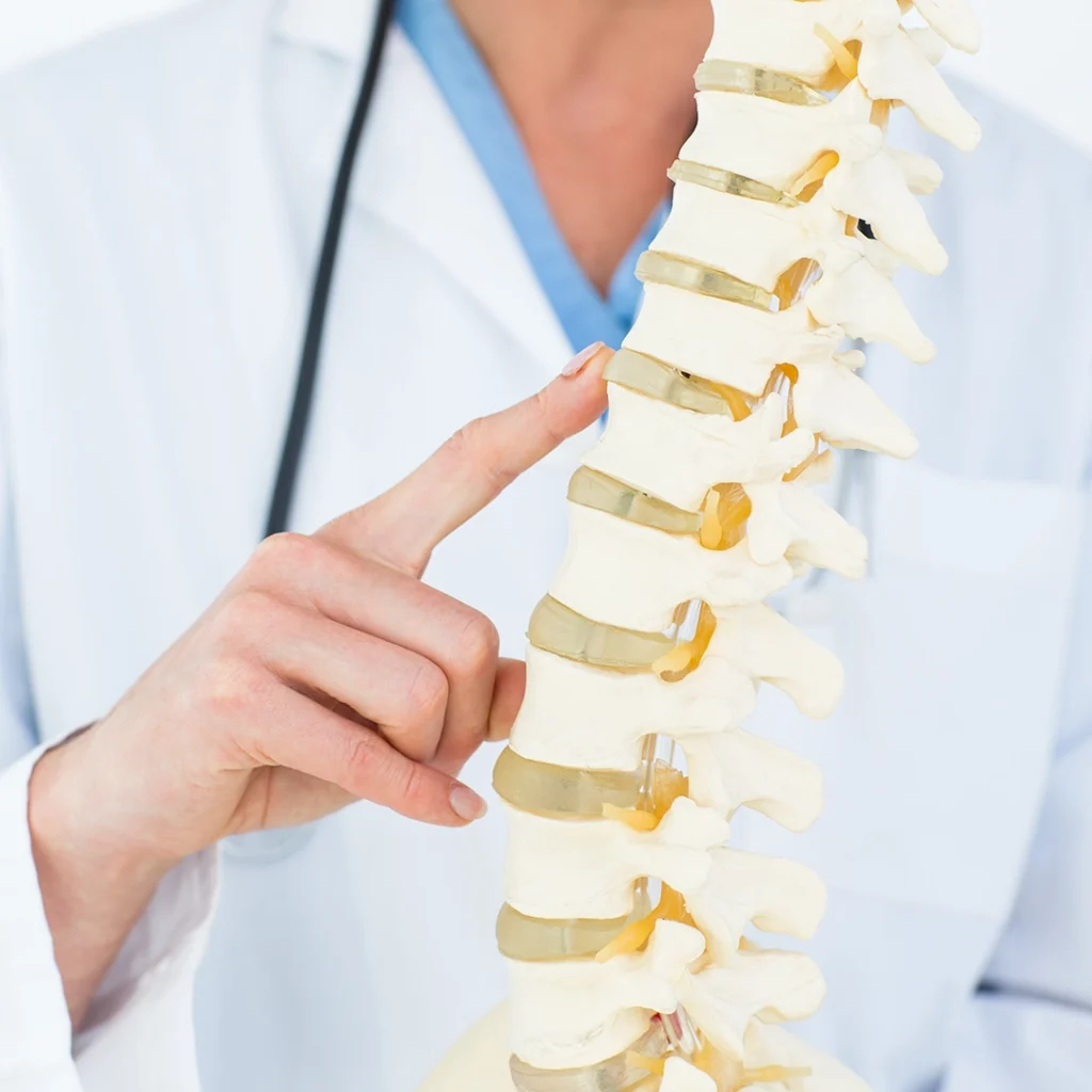
The knee skeleton consists mainly of:
The femur (bone located in the thigh, forming part of the hip and knee) articulates with the tibia (bone located in the lower leg in the inner area). In front of the femur and tibia is the patella (a flat bone), which slides into a groove when the knee moves. The fibula (bone located parallel to the tibia in the outer area) does not form the knee joint but is essential for the insertion of muscles and ligaments.
Bone structures are protected by the cartilage of the knee which reduces the friction and wear that is inevitable over the years. When this same cartilage degenerates, we speak of knee arthrosis, one of the main causes leading to knee replacement.
The menisci are also structures made of cartilage. They are located on the upper part of the tibia, provide stability and cushion the stresses that the femur and our body produce on the tibia. Each knee has two menisci the medial meniscus and the lateral meniscus.
We can suffer meniscus injury from trauma or wear and tear.
The ligaments that give stability to the knee during our activities are:
- anterior cruciate ligament and posterior cruciate ligament, often the site of knee injuries or sprains, especially in athletes (ACL or ACL surgery)
- internal collateral ligament and external collateral ligament
The main muscles of the knee are:
- quadriceps: in the anterior portion of the thigh, which extends the knee and flexes thehip
- biceps femoris: in the posterior portion of the thigh, which in cooperation with semitendinosus and gracilis, works to flex the knee
Causes and objective of knee treatment
We know that knee pain is very common, but knee replacement is not always the solution; let's try to understand in which cases it is necessary to intervene.
The most common cause is joint damage, i.e. wear and tear or injury to the skeletal components of the knee. Such conditions, in their most severe forms, produce:
- pain and swelling
- joint stiffness
- reduced knee mobility
- difficulty in carrying out normal daily activities
Most frequent causes of knee replacement surgery
The most common causes of knee replacement surgery are:
Knee arthrosis
It is a degenerative disease of the cartilage that covers the femur and tibia. When the cartilage is damaged, friction and degeneration of the bone tissue increases, resulting in increased pain, swelling and limited mobility of the knee.
Rheumatoid arthritis
It is a chronic autoimmune disease that attacks the bone tissue of small joints. The immune system of sufferers, instead of protecting the body from viruses and bacteria, turns against it, causing inflammation of synovial tissue which, in turn, triggers chronic pain and degeneration of cartilage and then bone tissue.
Haemophilia
It is a genetic disorder that alters the common blood clotting systems causing continuous haemorrhaging, which mainly affects knees and ankles. The consequence is weakening of these joints and subsequent stiffness and wear.
Other less common causes are gout (uric acid accumulation that inflames joints), vascular necrosis, repeated trauma involving menisci or ligaments, bone dysplasia and congenital deformities (e.g. varus or valgus knee).
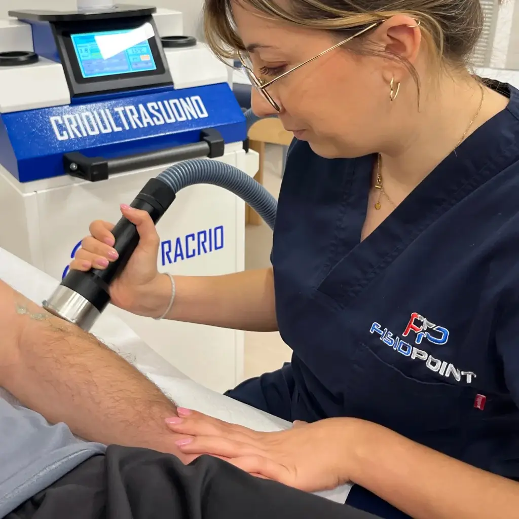
All these clinical conditions lead to progressive joint damage. This is why, in the first phase, one can intervene with the help of anti-inflammatory drugs and physiotherapy, which aims to reduce pain, inflammation, and joint stress by increasing the stability of the knee and re-educating movement. When, on the other hand, the symptoms become disabling, surgical treatment with knee replacements is necessary.
This approach mainly affects patients between 60 and 80 years of age. In young people, conservative treatment is preferred, since the prosthesis lasts about 15 to 20 years after which it needs to be revised or replaced by an even more invasive operation than the first. The longer the implantation of the prosthesis is delayed, the less likely it is to wear out and thus require revision.
Goals of knee replacement
The final objectives of the intervention are:
- decrease in pain
- increased knee mobility
- improvement of the patient's motor skills
- improvement of the patient's quality of life
Type of prosthesis and procedure
Depending on the extent of the joint damage and the age of the subject, the surgeon chooses between two types of prosthesis:
- more frequent total prosthesis, involves the replacement of a portion of the femur and tibia
- partial prosthesis, replacing the worn portion of a single joint head or femur or tibia
How total knee replacement surgery takes place
It is the most frequently chosen type of surgery by surgeons. The operation takes place under general or epidural anaesthesia and can last between one and three hours. We distinguish three phases:
- engraving
- removal of damaged bone portions
- replacement with prosthesis
The incision is vertical in line with the patella, which is then moved to completely clear the operating field. The damaged ends of the femur and tibia are removed and replaced with metal plates (curved for the femur and flat for the tibia) cemented to the bone. If the kneecap is also worn, its inner portion is replaced. A plastic insert is placed between the metal plates, which allows the surfaces to glide during knee movements and acts as cartilage to reduce friction between the parts. Once the implantation of the prosthesis is complete, the incision is closed and sutured.
How partial knee replacement surgery takes place
The operation follows the same steps as total joint replacement. The difference lies in the incision, which is smaller and less invasive, and in the replacement of the damaged portion of a single joint head or tibia or femur. The healing time is shorter as it is a less complex operation, but the partial prosthesis has disadvantages:
- the bone segment not replaced during surgery will wear out over the years and will need to be reworked
- the lifespan of partial dentures (10-15 years) is shorter than that of full dentures (15-20 years)
These conditions lead the surgeon to choose total prosthesis in most cases.
How to prepare for surgery
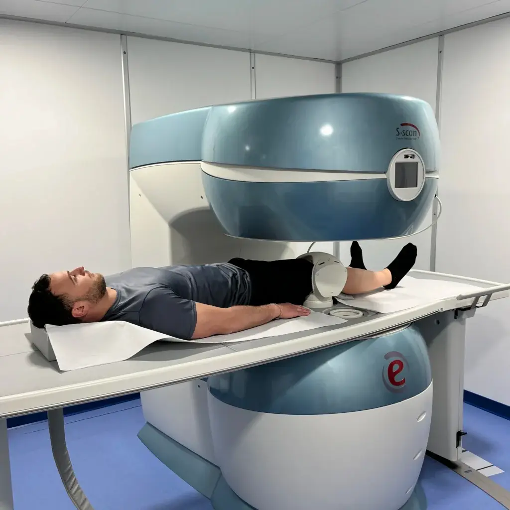
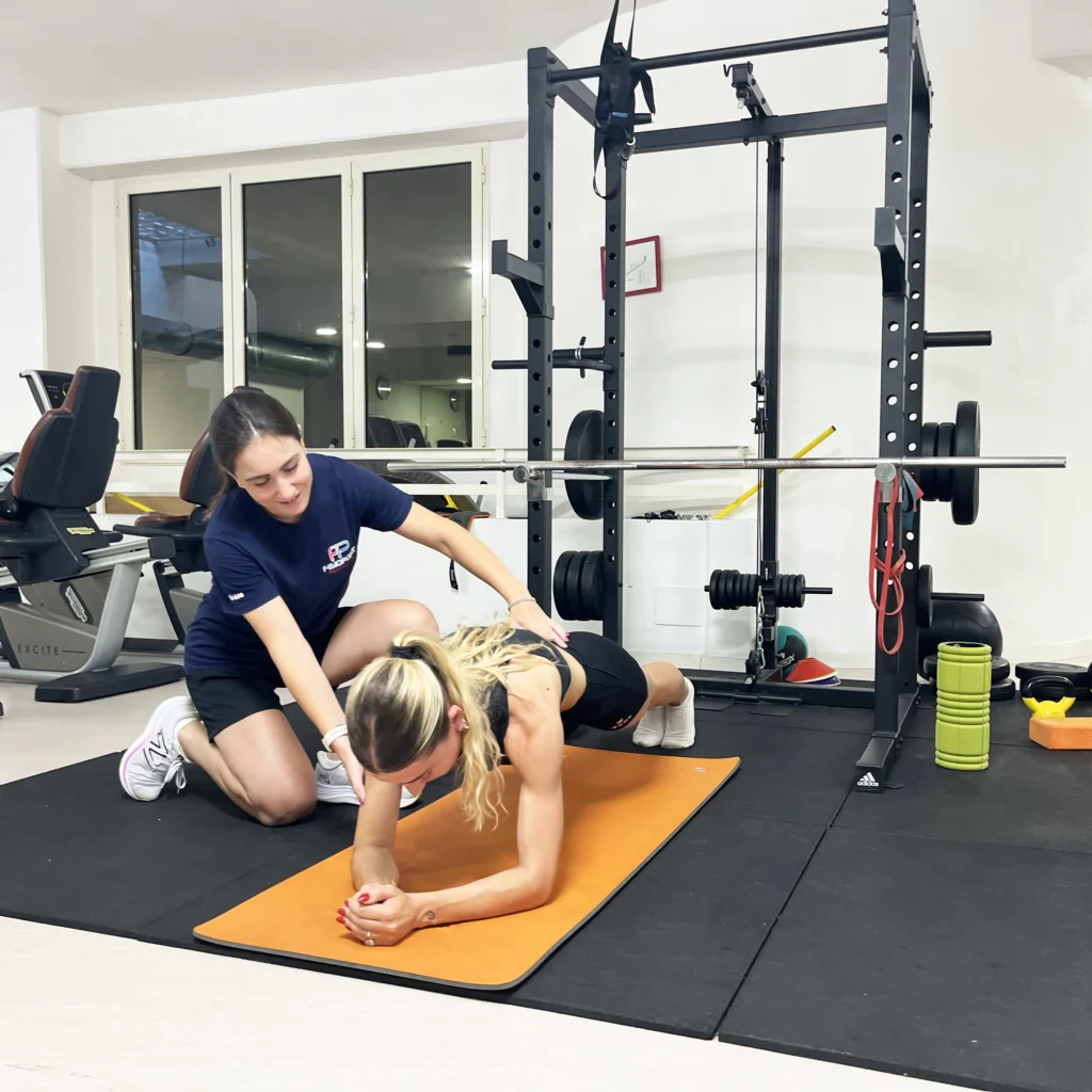
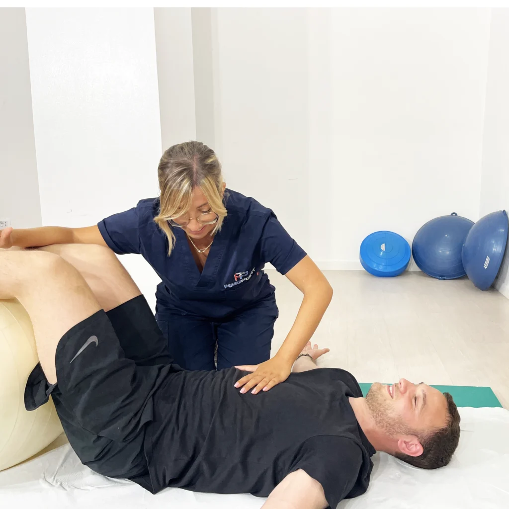
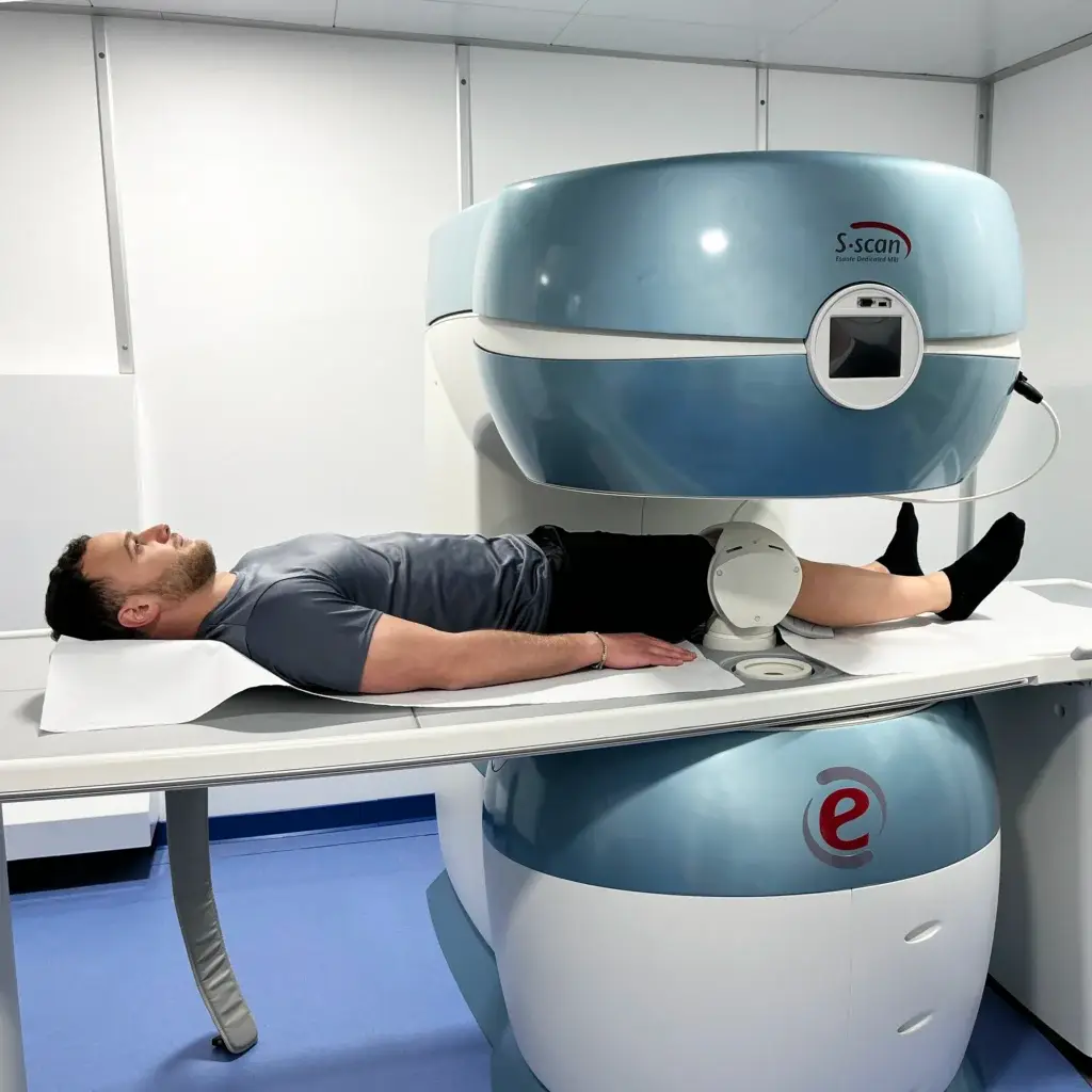
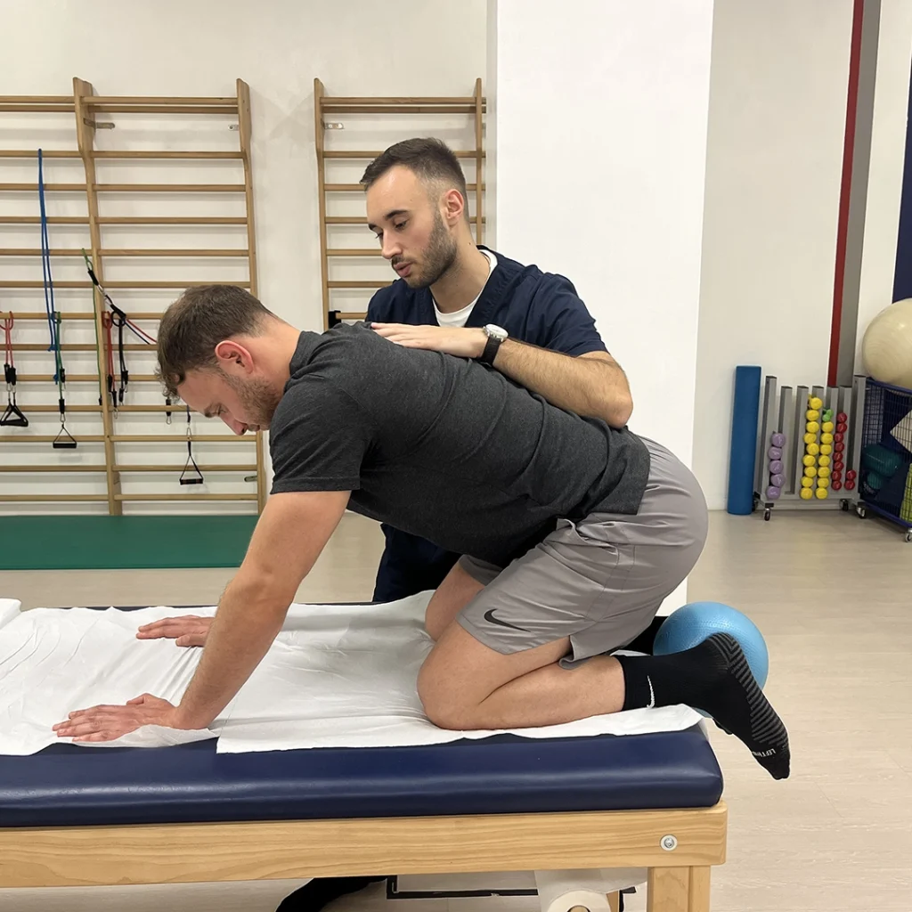
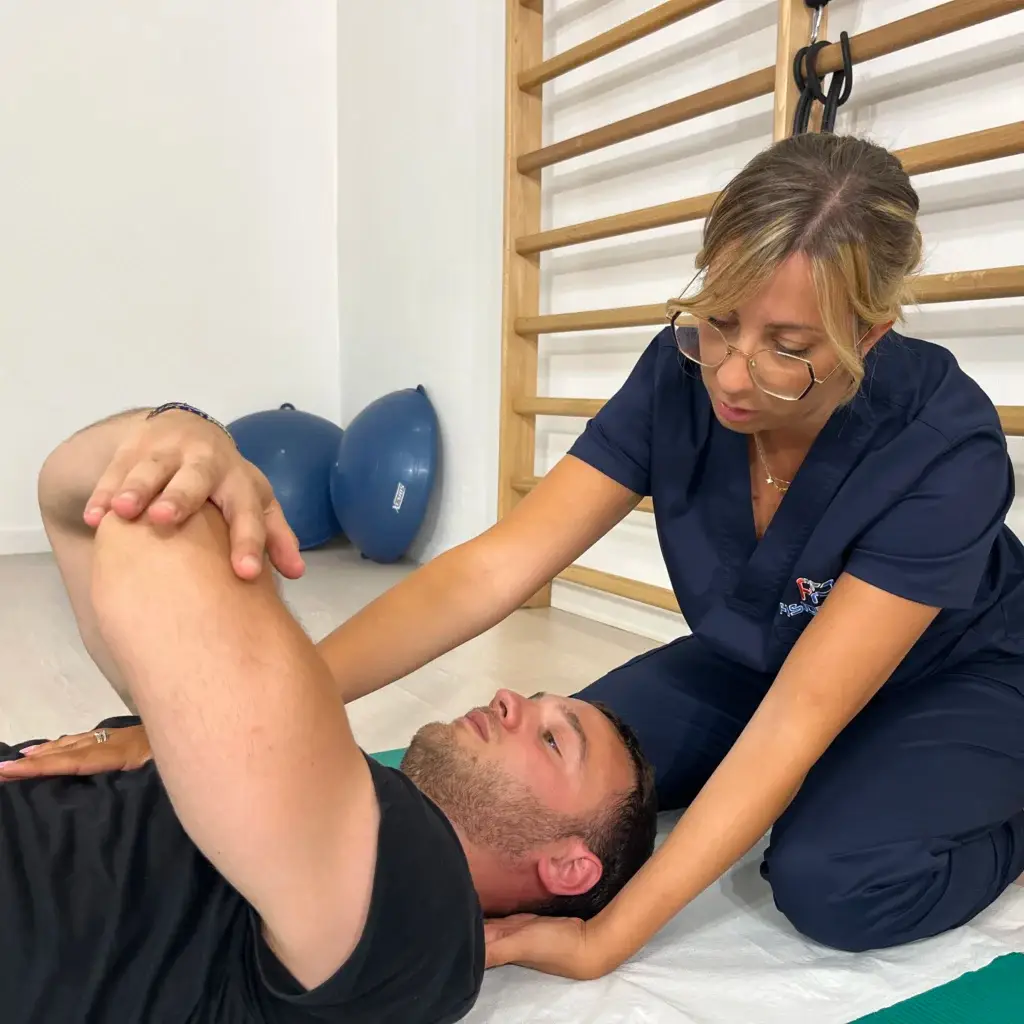
The pre-operative phase is often underestimated, instead it has a fundamental importance in shortening recovery time after surgery. It is necessary to prepare the body for the operation with physiotherapy, muscle stretching and strengthening exercises for the entire joint complex and core stability, without neglecting the upper limbs, which will be needed in the first few days for walking with Canadian canes. This is also the phase in which the patient's initial condition is studied, through instrumental examinations (rm, x-ray, electromyography) and the modalities of intervention are decided.
What to do after the operation
The post-operative phase is conditioned by several factors such as the patient's age, state of health
general, the type of prosthesis implanted, the preoperative physical condition.
The patient usually remains hospitalised for about a week, in order to monitor his general condition, but today, thanks to modern prostheses, studies confirm that rehabilitation can and should begin already after the first 24/48 h post-surgery. The patient will be able to stand and walk with the use of canes or a walker, which he/she will maintain for 3-6 weeks, the time needed for the repair of the muscular-ligament tissues. During this phase, the patient will receive heparin therapy to prevent thrombus formation due to the reduced walking. The overall recovery time, allowing the subject to resume his or her normal life, is approximately 3 months, during which time personal commitment and proper rehabilitation is essential. Total recovery occurs after 1-2 years.
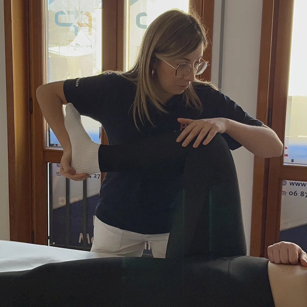
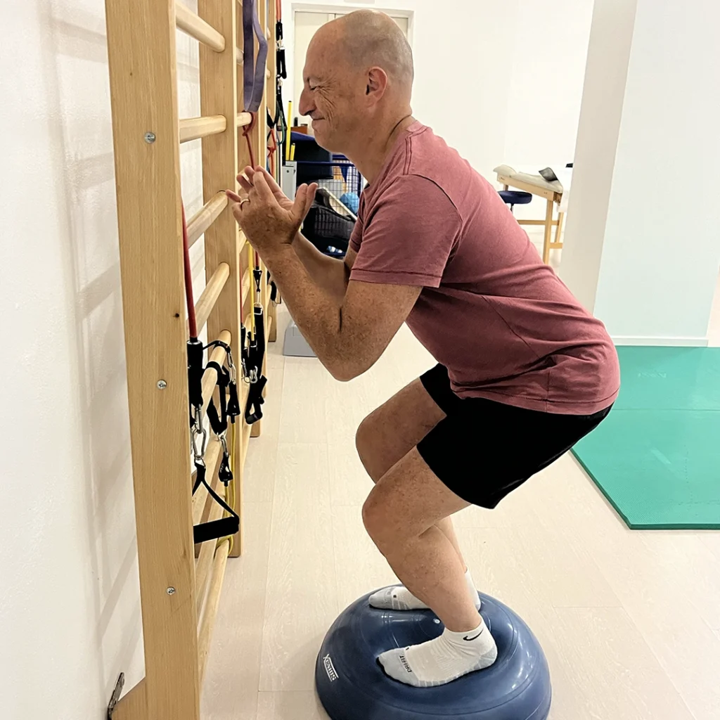
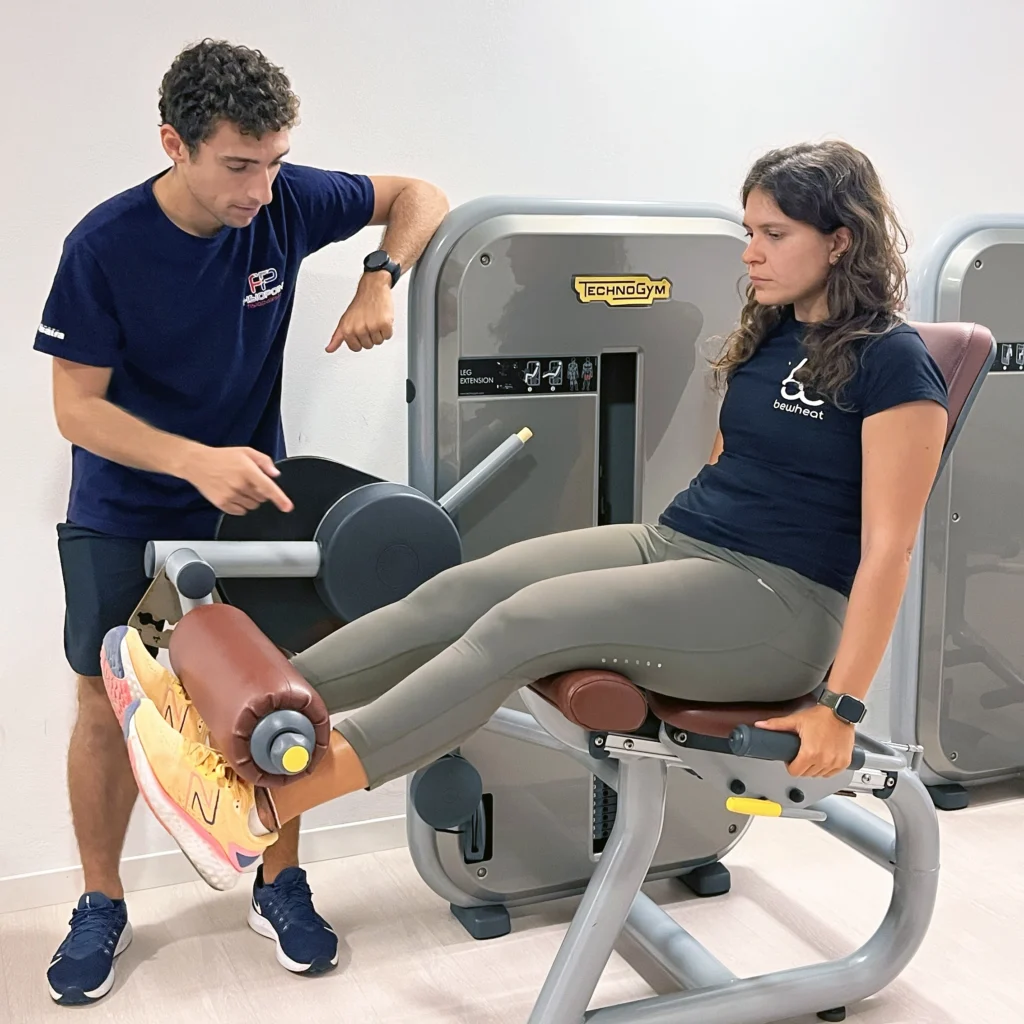
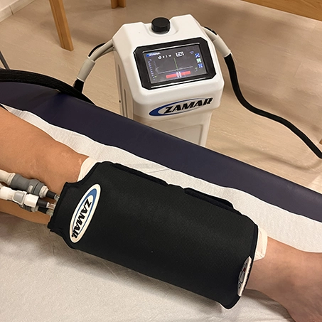
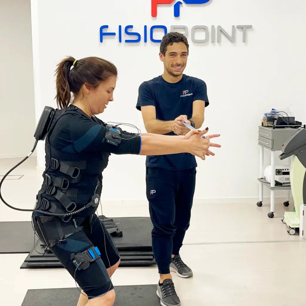
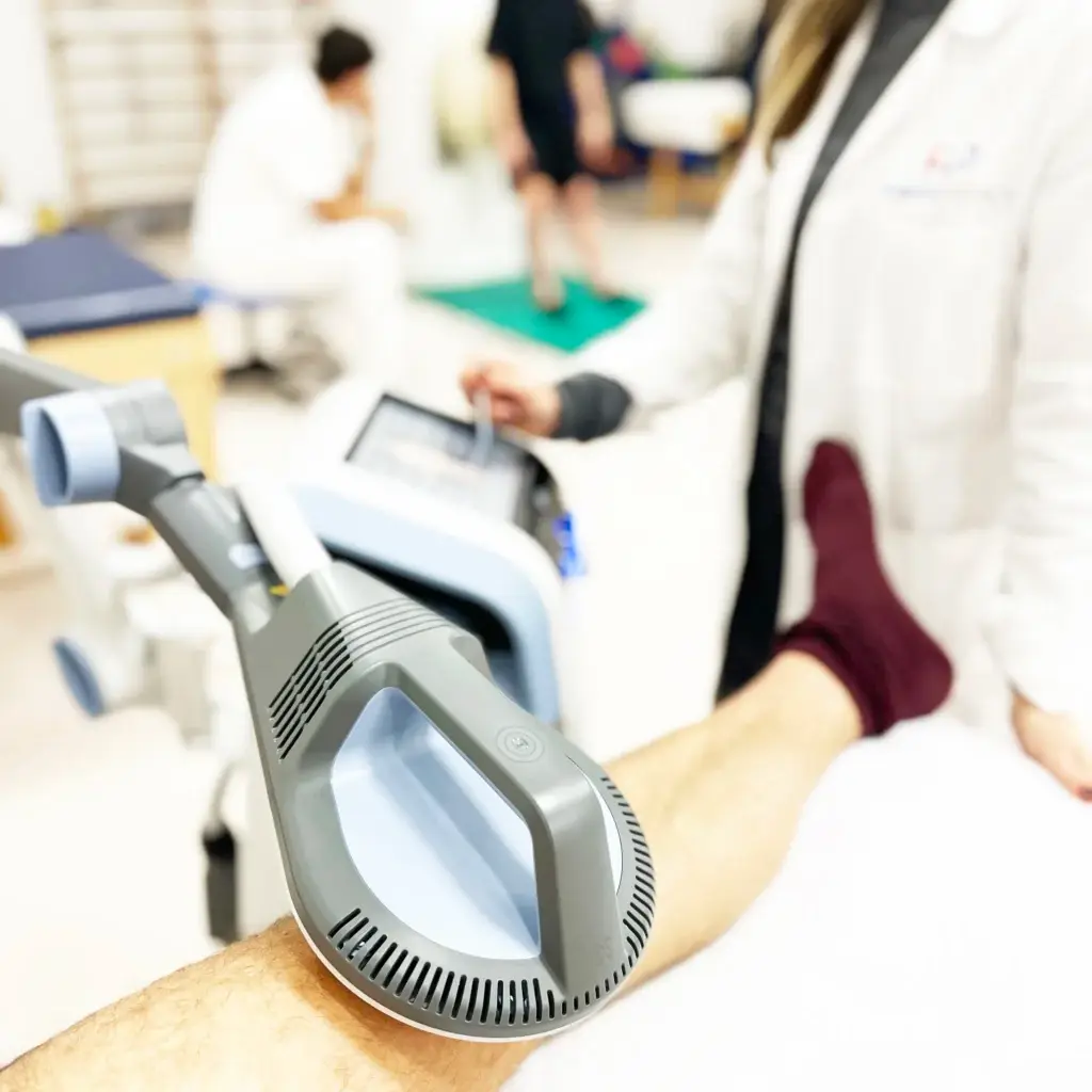
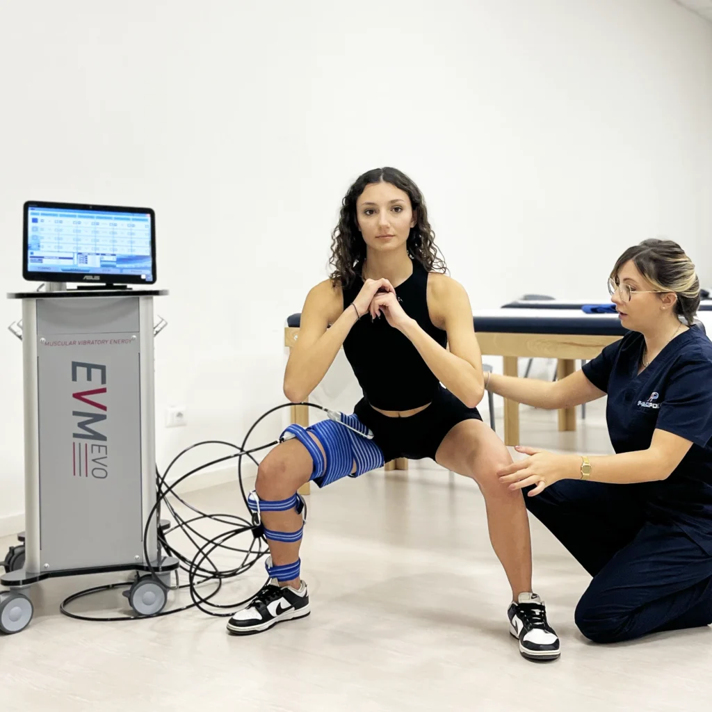
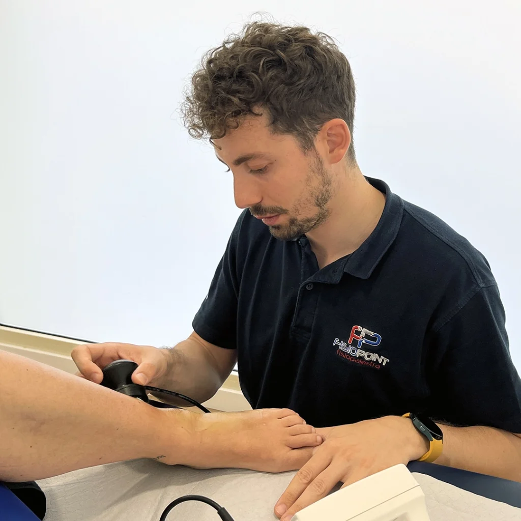
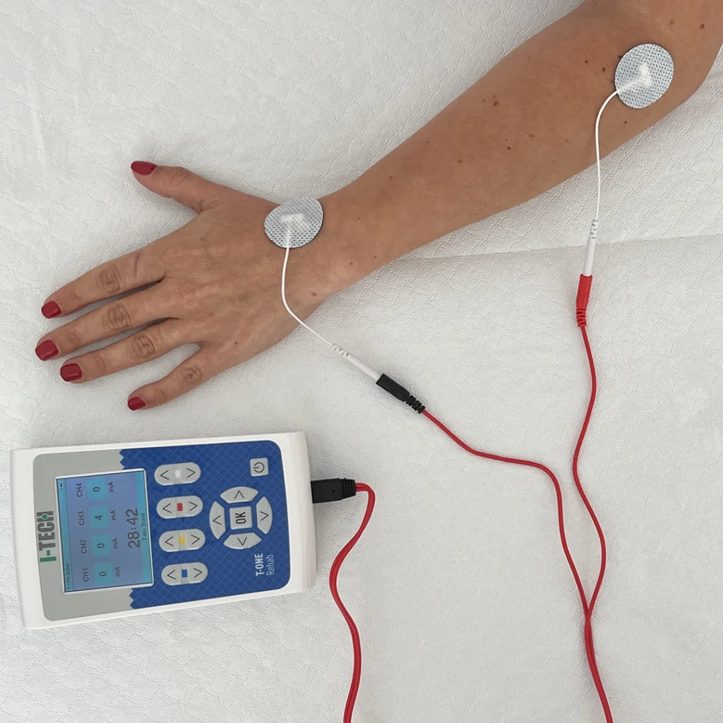
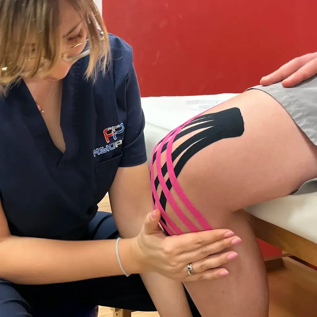
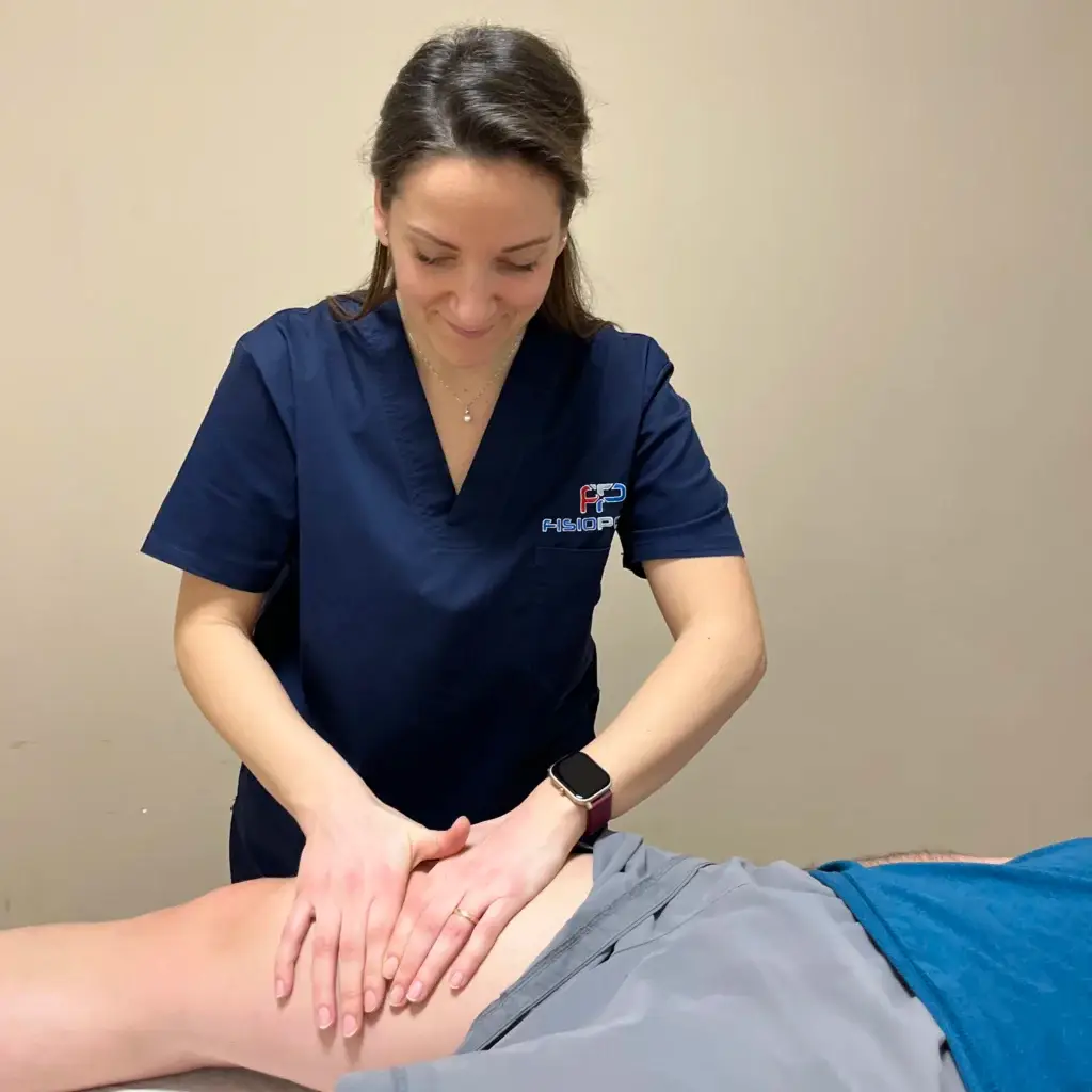
Physiotherapy is essential for:
- pain control
- recovery of knee articulation
- muscle strengthening of the joint complex and the whole body
- gait and proprioception re-education
- control of the emotional component: initial pain may discourage the patient but the support of a good physiotherapist help him overcome difficulties and achieve his goals
Text by Elisa Piccioni
Learn more by watching the videos on our channel YouTube









 English
 Italian