Anatomical prosthesis consists of replacing the joint with synthetic means that reconstruct the anatomy of the shoulder: humeral head (shaped like a portion of a sphere, similar in size to a golf ball) and scapular glenoid (concave surface, shallow and smaller than the head of the humerus).
The implantation of a anatomical prosthesis is effective when the rotator cuff is valid and is able to keep the prosthetic components centred while allowing correct movement
When anatomical prosthesis surgery is indicated
The conditions in which the implantation of an anatomical prosthesis is necessary are various: arthrosis, post-traumatic arthrosis, avascular necrosis, rheumatoid arthritis, fractures.
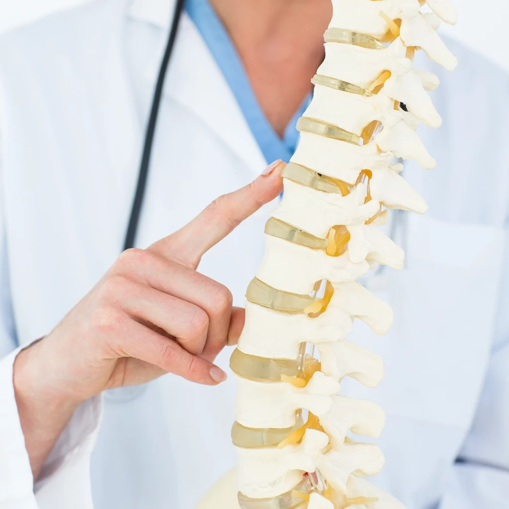
The most frequent of these is arthrosis, a chronic condition with an evolutionary character that involves a progressive lesion of the cartilage lining the joint surfaces, resulting in the disappearance of the joint space between the humerus and scapula, up to the deformation of the joint surfaces and the appearance of bony outgrowths called osteophytes. Sometimes it is an age-related process, others conditioned by previous damage, such as severe trauma or repeated dislocations of shoulderthus predisposing to an alteration of the biomechanics of the scapulohumeral girdle.
The arthrotic process is degenerative: initially, there is a decrease in glycosaminoglycans (molecules that, by binding to specific proteins, called proteoglycans, confer the particular characteristics of cushioning and therefore dispersion of mechanical stresses) and water, leading to the fragmentation of collagen fibres. On the surface, the cartilage crumbles until it disappears and exposes the subchondral bone (especially in the overload areas). Normally, cartilage is bluish white, moist and elastic, while in an arthrotic process it will appear opaque, rough, dry and softer and less elastic than normal.
The bone reacts with an overproduction of bone, particularly in the loaded areas, where it appears thickened; in the unloaded areas, along the perimeter of the articular surfaces, the blood vessels penetrate the cartilage giving rise to osteocartilaginous neoproduction, in an attempt to compensate for its loss: this results in the production of marginal osteophytes (beak-like bone rings), which remodel the articular heads, causing their progressive deformation.
The parameter that orients the surgeon to the implantation of an anatomical prosthesis is a high degree of arthrosis, which has not benefited from rehabilitation treatment or which is not compatible with this type of conservative approach from the outset.
Symptoms of arthrosis
The clinical picture of arthrosis is essentially characterised by two symptoms: pain and a progressive decrease in joint movement.
The pain has a gradual and aggravating evolution, is deep and generalised, arises at the beginning of movement, improves with rest, but in advanced stages is such that it prevents sleep.
From the point of view of mobility, in the early stages of arthrosis, passive range of motion is preserved, while active range of motion is painful; in the advanced stages, both passive and active range of motion are impossible, with the presence of crackling and rattling due to irregularities in the joint surfaces, and pain is not tolerated.
Arthrosis diagnosis
Confirmation of the above picture is carried out on the basis of clinical signs and symptoms and instrumental findings.
Useful instrumental investigations are:
- radiographic examination (true antero-posterior projection): is used to assess bone structures, relationships, joint surfaces and spaces, and soft tissue calcifications;
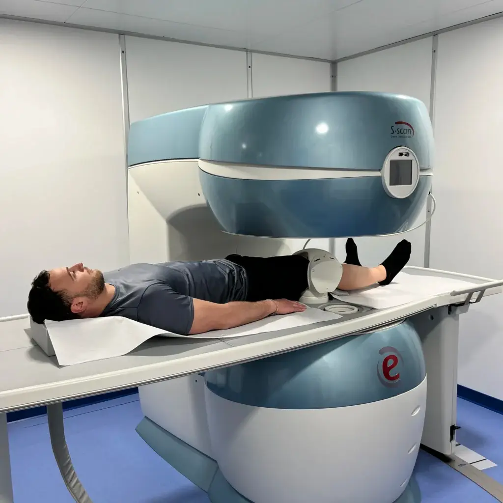
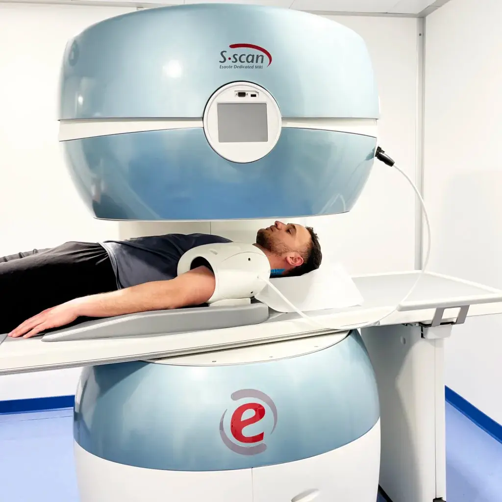
- magnetic resonance imagingto assess the state of the rotator cuff;
- CT scan (especially in the pre-operative phase): to guide the surgeon in choosing the type of implant.
Post-surgical rehabilitation for shoulder prosthesis
Following the implantation of the anatomical prosthesis, the patient must wear a brace that will position the arm in abduction and neutral rotation, for approximately three to four weeks; after the first 48/72 hours, the brace can begin to be removed for light activities and personal hygiene, wearing it instead during the night (unless otherwise indicated) and in all activities where it is easy to lose control of the limb.
Right from the start, it will be possible for the patient, on the therapist's instructions, to perform scapulothoracic recruitment movements, even while wearing the same brace, as well as extension and flexion movements of elbow.
Shoulder rehabilitation therapy
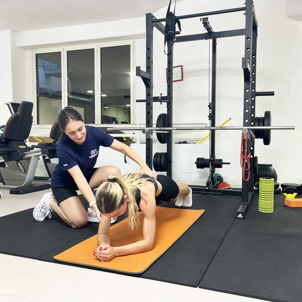
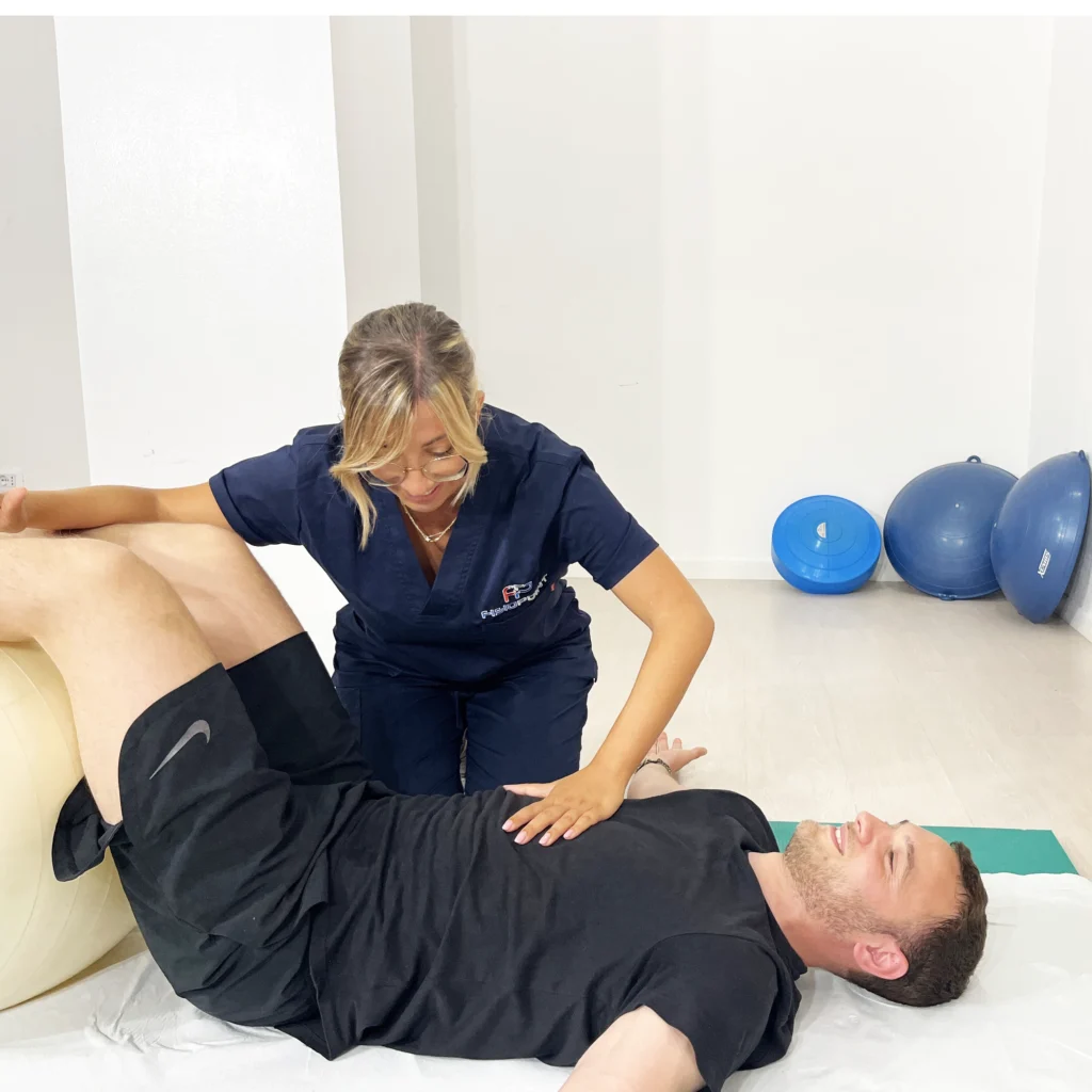
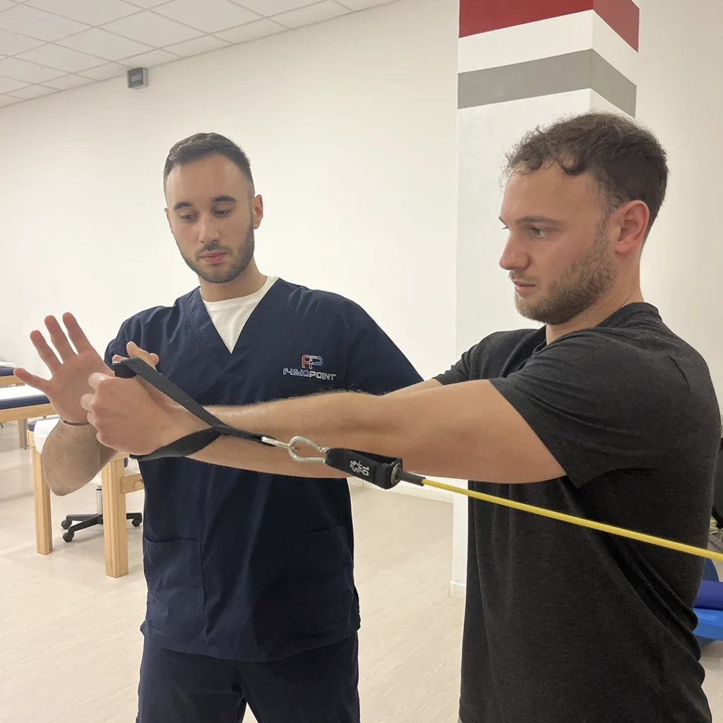
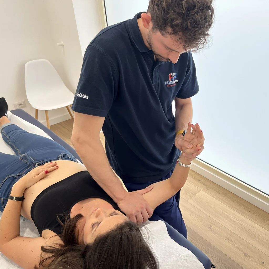
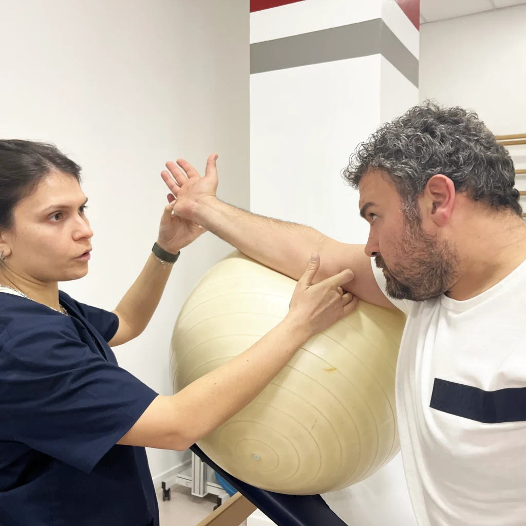
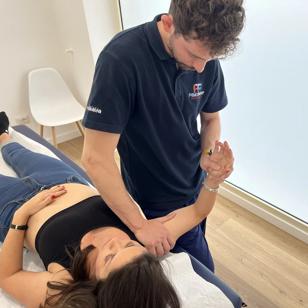
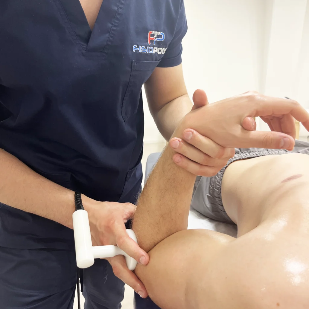
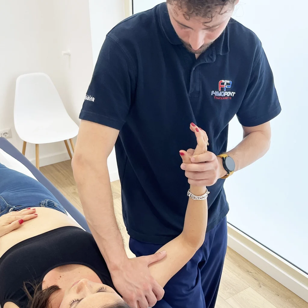
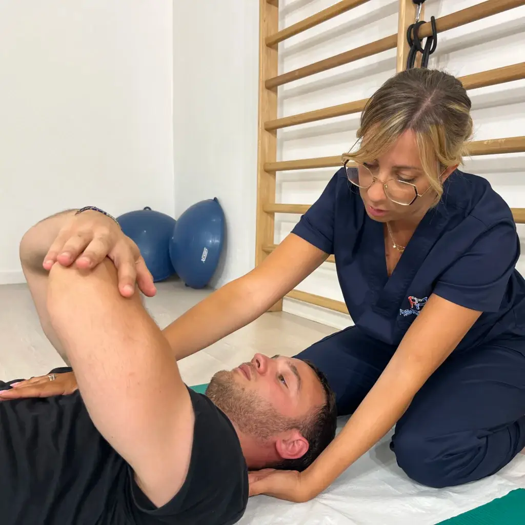
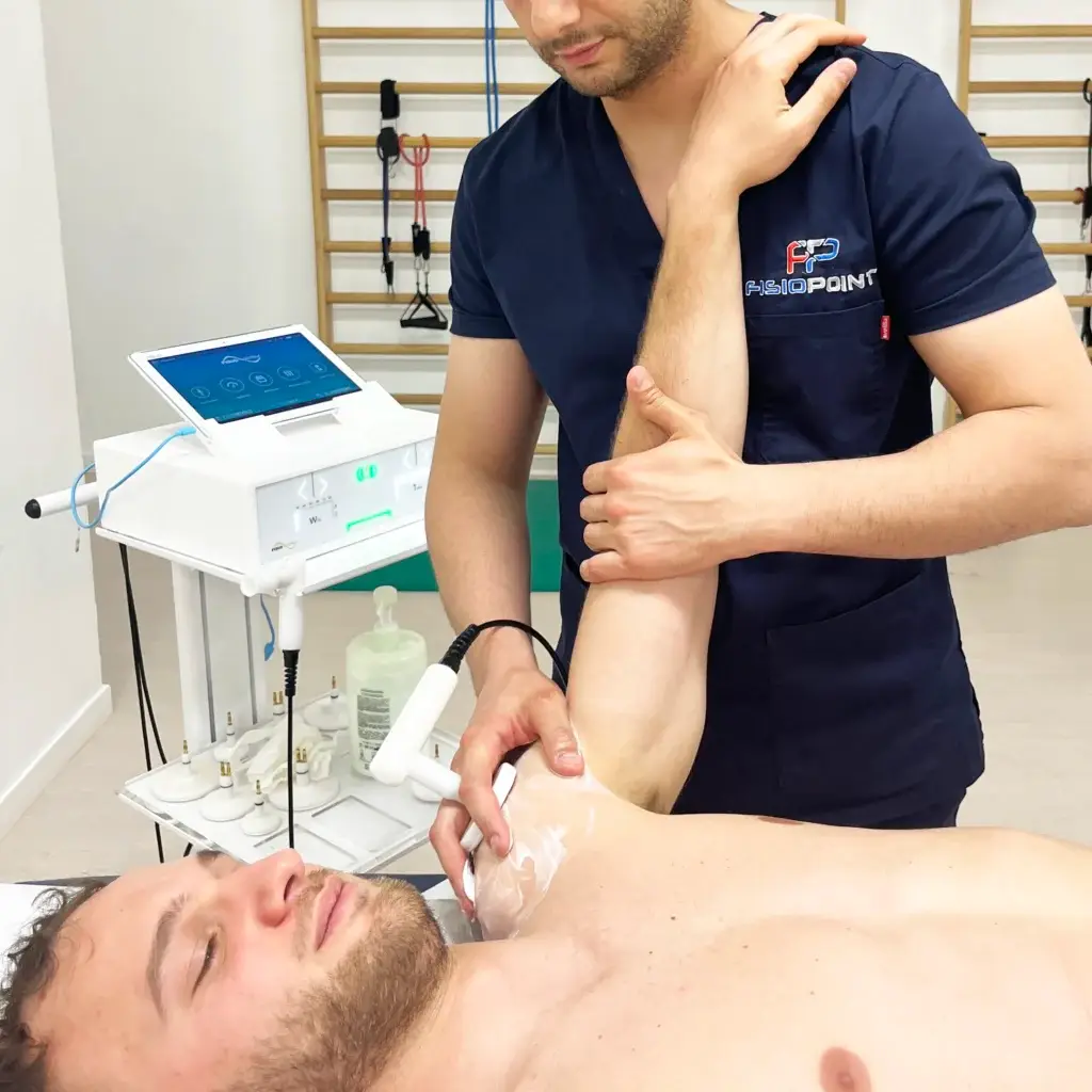
Rehabilitation therapyundertaken after the initial period of immobility in a brace, is a process of gradual recovery of mobility and strength of the shoulder, with the aim of regaining function as quickly as possible. In fact, in the first 6 weeks after removal of the brace, the aim is to recover full passive articularity, without pain, and active function in the 0-90° range; In this treatment window, the physiotherapist will have to balance the work of progressive muscular recruitment and that of gradual lengthening, while respecting the biological healing times (one of the surgical gestures performed is that of the tenotomy of the subscapularis, i.e., its incision 1 cm from the bony insertion and subsequent repair, the latter being a reason that must guide towards caution in internal rotation movements).
Improvement of pain
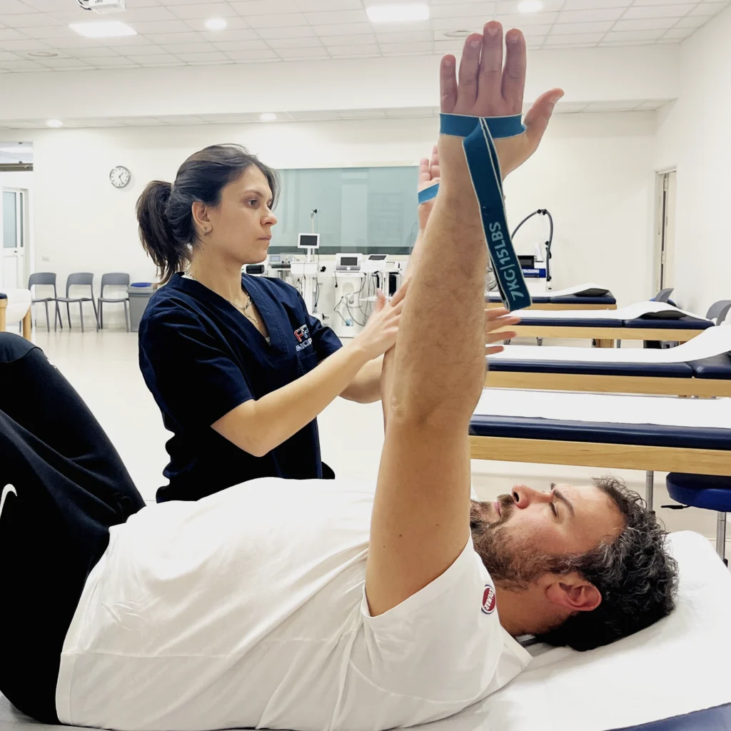
The progression of rehabilitation treatment in the second phase should aim at increasing activation of the rotator cuff muscles and increasing the active range of motion.
Reduction of pain and improvement of limb function after prosthetic implantation are achievable goals in more than 90% of cases.
The return to work will be agreed with the surgeon, depending on the type of task performed: the prosthesis is not suitable for heavy work, so it will be useful to readjust specific gestures to avoid premature wear of the prosthetic implant.
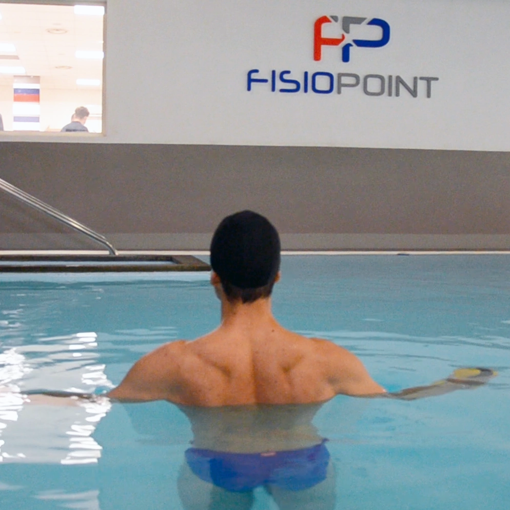
With the exception of traumatic cases, in all chronic diseases, where surgery can be planned well in advance, a preventive rehabilitation programme is recommended, so as to arrive at the day of surgery in the best possible condition. A programme of exercises of mobility and strengthening, pain arch excluded, associated, where possible, with cycles of hydrokinesiotherapyare a valuable support while waiting for the intervention.
Text by Elisa Piccioni
Learn more by watching the videos on our channel YouTube




 English
 Italian